Magnesium »
PDB 1ofh-1oyj »
1oix »
Magnesium in PDB 1oix: X-Ray Structure of the Small G Protein RAB11A in Complex with Gdp and Pi
Protein crystallography data
The structure of X-Ray Structure of the Small G Protein RAB11A in Complex with Gdp and Pi, PDB code: 1oix
was solved by
S.Pasqualato,
F.Senic-Matuglia,
L.Renault,
B.Goud,
J.Salamero,
J.Cherfils,
with X-Ray Crystallography technique. A brief refinement statistics is given in the table below:
| Resolution Low / High (Å) | 30 / 1.7 |
| Space group | I 4 2 2 |
| Cell size a, b, c (Å), α, β, γ (°) | 74.077, 74.077, 124.905, 90.00, 90.00, 90.00 |
| R / Rfree (%) | 20.1 / 22.9 |
Other elements in 1oix:
The structure of X-Ray Structure of the Small G Protein RAB11A in Complex with Gdp and Pi also contains other interesting chemical elements:
| Chlorine | (Cl) | 1 atom |
Magnesium Binding Sites:
The binding sites of Magnesium atom in the X-Ray Structure of the Small G Protein RAB11A in Complex with Gdp and Pi
(pdb code 1oix). This binding sites where shown within
5.0 Angstroms radius around Magnesium atom.
In total only one binding site of Magnesium was determined in the X-Ray Structure of the Small G Protein RAB11A in Complex with Gdp and Pi, PDB code: 1oix:
In total only one binding site of Magnesium was determined in the X-Ray Structure of the Small G Protein RAB11A in Complex with Gdp and Pi, PDB code: 1oix:
Magnesium binding site 1 out of 1 in 1oix
Go back to
Magnesium binding site 1 out
of 1 in the X-Ray Structure of the Small G Protein RAB11A in Complex with Gdp and Pi
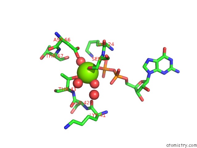
Mono view
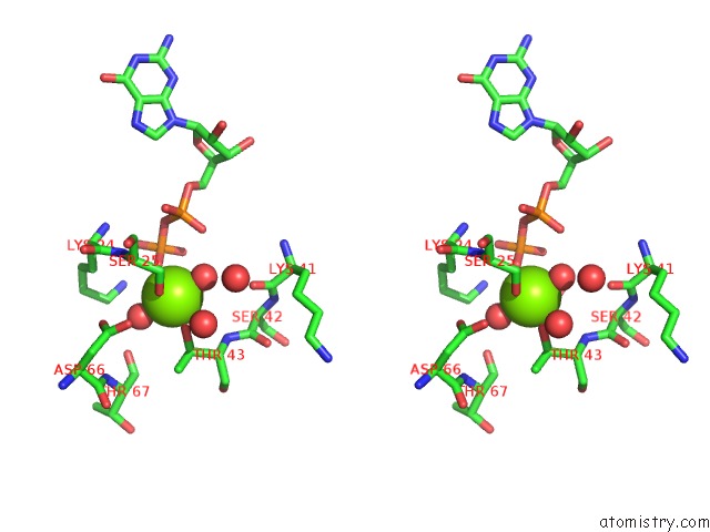
Stereo pair view
Mono view
Stereo pair view
A full contact list of Magnesium with other atoms in the Mg binding
site number 1 of X-Ray Structure of the Small G Protein RAB11A in Complex with Gdp and Pi within 5.0Å range:
|
Reference:
S.Pasqualato,
J.Cherfils.
Crystallographic Evidence For Substrate-Assisted Gtp Hydrolysis By A Small Gtp Binding Protein Structure V. 13 533 2005.
ISSN: ISSN 0969-2126
PubMed: 15837192
DOI: 10.1016/J.STR.2005.01.014
Page generated: Tue Aug 13 10:40:06 2024
ISSN: ISSN 0969-2126
PubMed: 15837192
DOI: 10.1016/J.STR.2005.01.014
Last articles
K in 4D7NK in 4CN5
K in 4CZB
K in 4D4T
K in 4CBB
K in 4CHI
K in 4CN0
K in 4CHW
K in 4CHV
K in 4CFS