Magnesium »
PDB 1ofh-1oyj »
1ol5 »
Magnesium in PDB 1ol5: Structure of Aurora-A 122-403, Phosphorylated on THR287, THR288 and Bound to TPX2 1-43
Enzymatic activity of Structure of Aurora-A 122-403, Phosphorylated on THR287, THR288 and Bound to TPX2 1-43
All present enzymatic activity of Structure of Aurora-A 122-403, Phosphorylated on THR287, THR288 and Bound to TPX2 1-43:
2.7.1.37;
2.7.1.37;
Protein crystallography data
The structure of Structure of Aurora-A 122-403, Phosphorylated on THR287, THR288 and Bound to TPX2 1-43, PDB code: 1ol5
was solved by
R.Bayliss,
E.Conti,
with X-Ray Crystallography technique. A brief refinement statistics is given in the table below:
| Resolution Low / High (Å) | 40 / 2.5 |
| Space group | P 21 21 21 |
| Cell size a, b, c (Å), α, β, γ (°) | 59.630, 81.720, 83.050, 90.00, 90.00, 90.00 |
| R / Rfree (%) | 19.4 / 25.2 |
Magnesium Binding Sites:
The binding sites of Magnesium atom in the Structure of Aurora-A 122-403, Phosphorylated on THR287, THR288 and Bound to TPX2 1-43
(pdb code 1ol5). This binding sites where shown within
5.0 Angstroms radius around Magnesium atom.
In total 3 binding sites of Magnesium where determined in the Structure of Aurora-A 122-403, Phosphorylated on THR287, THR288 and Bound to TPX2 1-43, PDB code: 1ol5:
Jump to Magnesium binding site number: 1; 2; 3;
In total 3 binding sites of Magnesium where determined in the Structure of Aurora-A 122-403, Phosphorylated on THR287, THR288 and Bound to TPX2 1-43, PDB code: 1ol5:
Jump to Magnesium binding site number: 1; 2; 3;
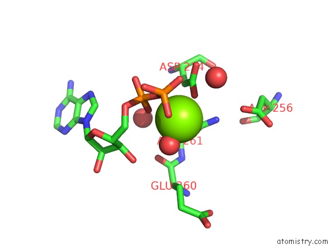
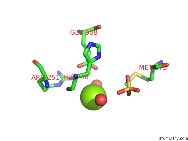
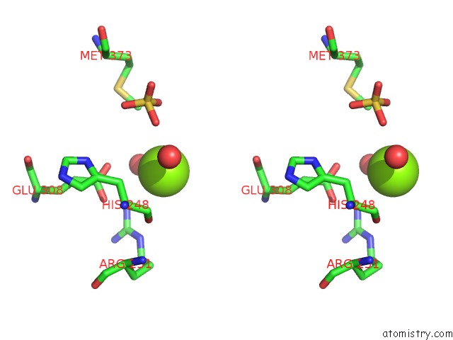
Magnesium binding site 1 out of 3 in 1ol5
Go back to
Magnesium binding site 1 out
of 3 in the Structure of Aurora-A 122-403, Phosphorylated on THR287, THR288 and Bound to TPX2 1-43
Mono view
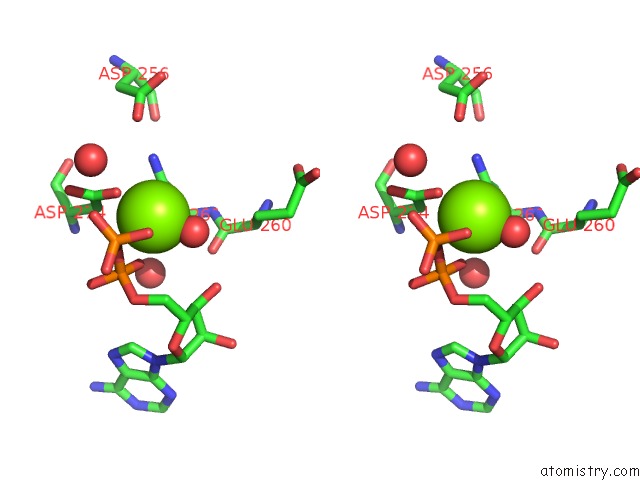
Stereo pair view
Mono view
Stereo pair view
A full contact list of Magnesium with other atoms in the Mg binding
site number 1 of Structure of Aurora-A 122-403, Phosphorylated on THR287, THR288 and Bound to TPX2 1-43 within 5.0Å range:
|
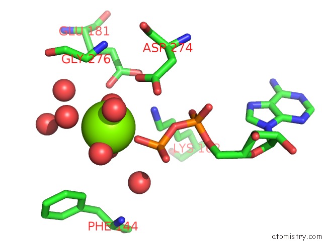
Magnesium binding site 2 out of 3 in 1ol5
Go back to
Magnesium binding site 2 out
of 3 in the Structure of Aurora-A 122-403, Phosphorylated on THR287, THR288 and Bound to TPX2 1-43
Mono view
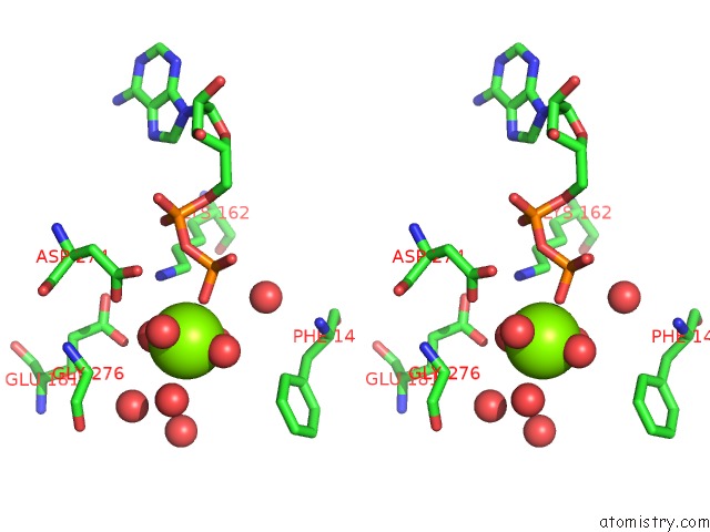
Stereo pair view
Mono view
Stereo pair view
A full contact list of Magnesium with other atoms in the Mg binding
site number 2 of Structure of Aurora-A 122-403, Phosphorylated on THR287, THR288 and Bound to TPX2 1-43 within 5.0Å range:
|
Magnesium binding site 3 out of 3 in 1ol5
Go back to
Magnesium binding site 3 out
of 3 in the Structure of Aurora-A 122-403, Phosphorylated on THR287, THR288 and Bound to TPX2 1-43
Mono view
Stereo pair view
Mono view
Stereo pair view
A full contact list of Magnesium with other atoms in the Mg binding
site number 3 of Structure of Aurora-A 122-403, Phosphorylated on THR287, THR288 and Bound to TPX2 1-43 within 5.0Å range:
|
Reference:
R.Bayliss,
T.Sardon,
I.Vernos,
E.Conti.
Structural Basis of Aurora-A Activation By TPX2 at the Mitotic Spindle Mol.Cell V. 12 851 2003.
ISSN: ISSN 1097-2765
PubMed: 14580337
DOI: 10.1016/S1097-2765(03)00392-7
Page generated: Tue Aug 13 10:40:32 2024
ISSN: ISSN 1097-2765
PubMed: 14580337
DOI: 10.1016/S1097-2765(03)00392-7
Last articles
K in 9CZMK in 9CZK
K in 9CZH
K in 9CZJ
K in 9CTU
K in 9CIY
K in 9CEN
K in 9CKO
K in 9CKP
K in 9CB5