Magnesium »
PDB 1ofh-1oyj »
1oxh »
Magnesium in PDB 1oxh: The Crystal Structure of Beta-Ketoacyl-[Acyl Carrier Protein] Synthase II From Streptococcus Pneumoniae, Triclinic Form
Enzymatic activity of The Crystal Structure of Beta-Ketoacyl-[Acyl Carrier Protein] Synthase II From Streptococcus Pneumoniae, Triclinic Form
All present enzymatic activity of The Crystal Structure of Beta-Ketoacyl-[Acyl Carrier Protein] Synthase II From Streptococcus Pneumoniae, Triclinic Form:
2.3.1.41;
2.3.1.41;
Protein crystallography data
The structure of The Crystal Structure of Beta-Ketoacyl-[Acyl Carrier Protein] Synthase II From Streptococcus Pneumoniae, Triclinic Form, PDB code: 1oxh
was solved by
A.C.Price,
C.O.Rock,
S.W.White,
with X-Ray Crystallography technique. A brief refinement statistics is given in the table below:
| Resolution Low / High (Å) | 29.19 / 2.09 |
| Space group | P 1 |
| Cell size a, b, c (Å), α, β, γ (°) | 61.520, 71.646, 96.100, 89.83, 83.09, 69.15 |
| R / Rfree (%) | 20.8 / 23.5 |
Magnesium Binding Sites:
The binding sites of Magnesium atom in the The Crystal Structure of Beta-Ketoacyl-[Acyl Carrier Protein] Synthase II From Streptococcus Pneumoniae, Triclinic Form
(pdb code 1oxh). This binding sites where shown within
5.0 Angstroms radius around Magnesium atom.
In total 4 binding sites of Magnesium where determined in the The Crystal Structure of Beta-Ketoacyl-[Acyl Carrier Protein] Synthase II From Streptococcus Pneumoniae, Triclinic Form, PDB code: 1oxh:
Jump to Magnesium binding site number: 1; 2; 3; 4;
In total 4 binding sites of Magnesium where determined in the The Crystal Structure of Beta-Ketoacyl-[Acyl Carrier Protein] Synthase II From Streptococcus Pneumoniae, Triclinic Form, PDB code: 1oxh:
Jump to Magnesium binding site number: 1; 2; 3; 4;
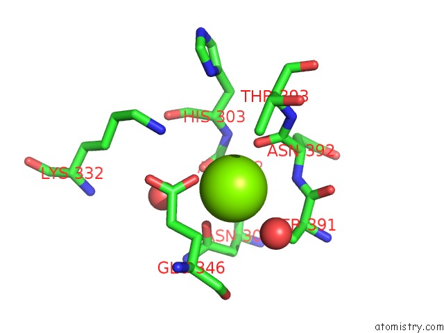
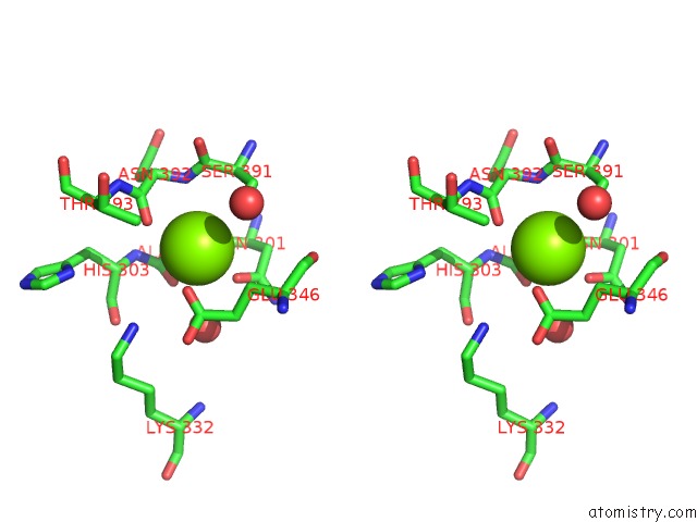
Magnesium binding site 1 out of 4 in 1oxh
Go back to
Magnesium binding site 1 out
of 4 in the The Crystal Structure of Beta-Ketoacyl-[Acyl Carrier Protein] Synthase II From Streptococcus Pneumoniae, Triclinic Form
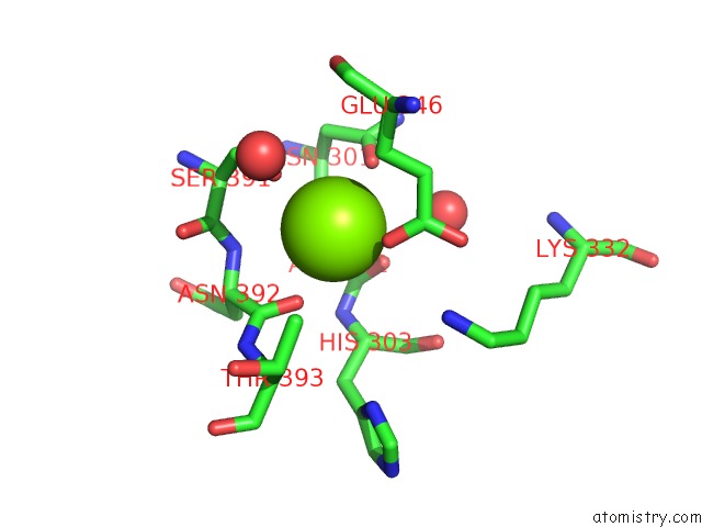
Mono view
Stereo pair view
Mono view
Stereo pair view
A full contact list of Magnesium with other atoms in the Mg binding
site number 1 of The Crystal Structure of Beta-Ketoacyl-[Acyl Carrier Protein] Synthase II From Streptococcus Pneumoniae, Triclinic Form within 5.0Å range:
|
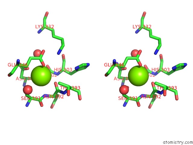
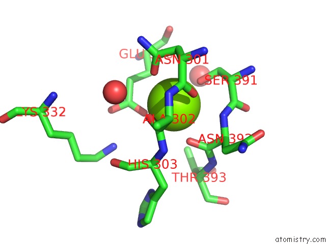
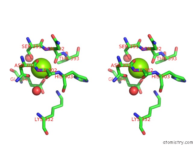
Magnesium binding site 2 out of 4 in 1oxh
Go back to
Magnesium binding site 2 out
of 4 in the The Crystal Structure of Beta-Ketoacyl-[Acyl Carrier Protein] Synthase II From Streptococcus Pneumoniae, Triclinic Form
Mono view
Stereo pair view
Mono view
Stereo pair view
A full contact list of Magnesium with other atoms in the Mg binding
site number 2 of The Crystal Structure of Beta-Ketoacyl-[Acyl Carrier Protein] Synthase II From Streptococcus Pneumoniae, Triclinic Form within 5.0Å range:
|
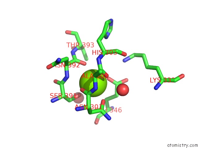
Magnesium binding site 3 out of 4 in 1oxh
Go back to
Magnesium binding site 3 out
of 4 in the The Crystal Structure of Beta-Ketoacyl-[Acyl Carrier Protein] Synthase II From Streptococcus Pneumoniae, Triclinic Form
Mono view
Stereo pair view
Mono view
Stereo pair view
A full contact list of Magnesium with other atoms in the Mg binding
site number 3 of The Crystal Structure of Beta-Ketoacyl-[Acyl Carrier Protein] Synthase II From Streptococcus Pneumoniae, Triclinic Form within 5.0Å range:
|
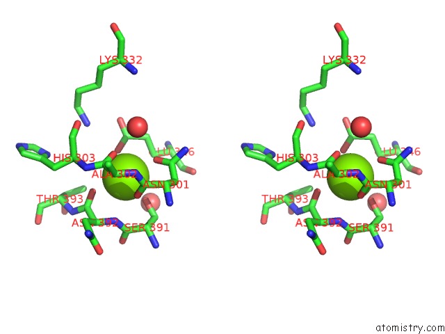
Magnesium binding site 4 out of 4 in 1oxh
Go back to
Magnesium binding site 4 out
of 4 in the The Crystal Structure of Beta-Ketoacyl-[Acyl Carrier Protein] Synthase II From Streptococcus Pneumoniae, Triclinic Form
Mono view
Stereo pair view
Mono view
Stereo pair view
A full contact list of Magnesium with other atoms in the Mg binding
site number 4 of The Crystal Structure of Beta-Ketoacyl-[Acyl Carrier Protein] Synthase II From Streptococcus Pneumoniae, Triclinic Form within 5.0Å range:
|
Reference:
A.C.Price,
C.O.Rock,
S.W.White.
The 1.3-Angstrom-Resolution Crystal Structure of Beta-Ketoacyl-Acyl Carrier Protein Synthase II From Streptococcus Pneumoniae. J.Bacteriol. V. 185 4136 2003.
ISSN: ISSN 0021-9193
PubMed: 12837788
DOI: 10.1128/JB.185.14.4136-4143.2003
Page generated: Tue Aug 13 10:44:19 2024
ISSN: ISSN 0021-9193
PubMed: 12837788
DOI: 10.1128/JB.185.14.4136-4143.2003
Last articles
Mg in 1HQMMg in 1HQC
Mg in 1HQ2
Mg in 1HQ1
Mg in 1HPM
Mg in 1HN1
Mg in 1HC8
Mg in 1HMV
Mg in 1HI0
Mg in 1HJK