Magnesium »
PDB 1q3b-1qc1 »
1q3b »
Magnesium in PDB 1q3b: Crystal Structure of the Dna Repair Enzyme Endonuclease-VIII (Nei) From E. Coli: the R252A Mutant at 2.05 Resolution.
Protein crystallography data
The structure of Crystal Structure of the Dna Repair Enzyme Endonuclease-VIII (Nei) From E. Coli: the R252A Mutant at 2.05 Resolution., PDB code: 1q3b
was solved by
G.Golan,
D.O.Zharkov,
H.Feinberg,
A.S.Fernandes,
E.I.Zaika,
J.H.Kycia,
A.P.Grollman,
G.Shoham,
with X-Ray Crystallography technique. A brief refinement statistics is given in the table below:
| Resolution Low / High (Å) | 29.21 / 2.05 |
| Space group | I 2 2 2 |
| Cell size a, b, c (Å), α, β, γ (°) | 57.722, 80.051, 169.311, 90.00, 90.00, 90.00 |
| R / Rfree (%) | 22.8 / 26 |
Other elements in 1q3b:
The structure of Crystal Structure of the Dna Repair Enzyme Endonuclease-VIII (Nei) From E. Coli: the R252A Mutant at 2.05 Resolution. also contains other interesting chemical elements:
| Zinc | (Zn) | 1 atom |
Magnesium Binding Sites:
The binding sites of Magnesium atom in the Crystal Structure of the Dna Repair Enzyme Endonuclease-VIII (Nei) From E. Coli: the R252A Mutant at 2.05 Resolution.
(pdb code 1q3b). This binding sites where shown within
5.0 Angstroms radius around Magnesium atom.
In total 2 binding sites of Magnesium where determined in the Crystal Structure of the Dna Repair Enzyme Endonuclease-VIII (Nei) From E. Coli: the R252A Mutant at 2.05 Resolution., PDB code: 1q3b:
Jump to Magnesium binding site number: 1; 2;
In total 2 binding sites of Magnesium where determined in the Crystal Structure of the Dna Repair Enzyme Endonuclease-VIII (Nei) From E. Coli: the R252A Mutant at 2.05 Resolution., PDB code: 1q3b:
Jump to Magnesium binding site number: 1; 2;
Magnesium binding site 1 out of 2 in 1q3b
Go back to
Magnesium binding site 1 out
of 2 in the Crystal Structure of the Dna Repair Enzyme Endonuclease-VIII (Nei) From E. Coli: the R252A Mutant at 2.05 Resolution.
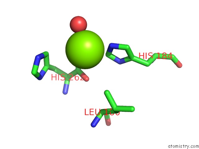
Mono view
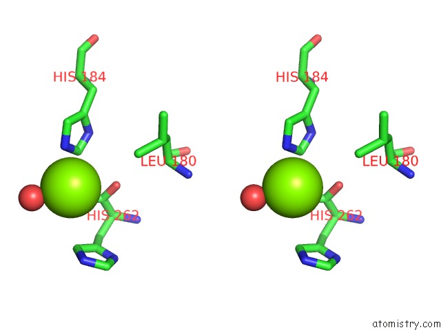
Stereo pair view
Mono view
Stereo pair view
A full contact list of Magnesium with other atoms in the Mg binding
site number 1 of Crystal Structure of the Dna Repair Enzyme Endonuclease-VIII (Nei) From E. Coli: the R252A Mutant at 2.05 Resolution. within 5.0Å range:
|
Magnesium binding site 2 out of 2 in 1q3b
Go back to
Magnesium binding site 2 out
of 2 in the Crystal Structure of the Dna Repair Enzyme Endonuclease-VIII (Nei) From E. Coli: the R252A Mutant at 2.05 Resolution.
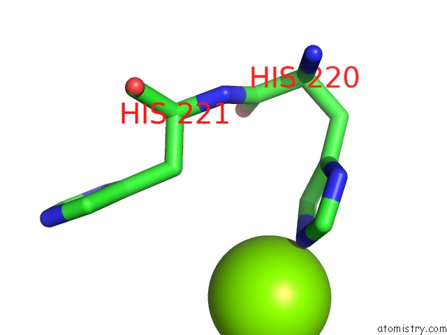
Mono view
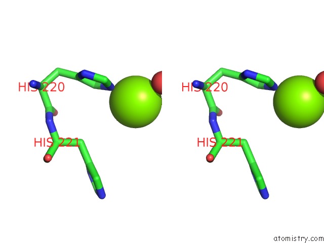
Stereo pair view
Mono view
Stereo pair view
A full contact list of Magnesium with other atoms in the Mg binding
site number 2 of Crystal Structure of the Dna Repair Enzyme Endonuclease-VIII (Nei) From E. Coli: the R252A Mutant at 2.05 Resolution. within 5.0Å range:
|
Reference:
G.Golan,
D.O.Zharkov,
H.Feinberg,
A.S.Fernandes,
E.I.Zaika,
J.H.Kycia,
A.P.Grollman,
G.Shoham.
Structure of the Uncomplexed Dna Repair Enzyme Endonuclease VIII Indicates Significant Interdomain Flexibility. Nucleic Acids Res. V. 33 5006 2005.
ISSN: ISSN 0305-1048
PubMed: 16145054
DOI: 10.1093/NAR/GKI796
Page generated: Sun Aug 10 02:27:53 2025
ISSN: ISSN 0305-1048
PubMed: 16145054
DOI: 10.1093/NAR/GKI796
Last articles
Mn in 2WOCMn in 2WOE
Mn in 2WZF
Mn in 2WQP
Mn in 2WOD
Mn in 2WJF
Mn in 2WHO
Mn in 2WJE
Mn in 2WJD
Mn in 2WHX