Magnesium »
PDB 1qsi-1rc5 »
1r6w »
Magnesium in PDB 1r6w: Crystal Structure of the K133R Mutant of O-Succinylbenzoate Synthase (Osbs) From Escherichia Coli. Complex with Shchc
Protein crystallography data
The structure of Crystal Structure of the K133R Mutant of O-Succinylbenzoate Synthase (Osbs) From Escherichia Coli. Complex with Shchc, PDB code: 1r6w
was solved by
V.A.Klenchin,
E.A.Taylor Ringia,
J.A.Gerlt,
I.Rayment,
with X-Ray Crystallography technique. A brief refinement statistics is given in the table below:
| Resolution Low / High (Å) | 30.00 / 1.62 |
| Space group | P 21 21 2 |
| Cell size a, b, c (Å), α, β, γ (°) | 72.200, 82.900, 56.200, 90.00, 90.00, 90.00 |
| R / Rfree (%) | 16.7 / 20 |
Magnesium Binding Sites:
The binding sites of Magnesium atom in the Crystal Structure of the K133R Mutant of O-Succinylbenzoate Synthase (Osbs) From Escherichia Coli. Complex with Shchc
(pdb code 1r6w). This binding sites where shown within
5.0 Angstroms radius around Magnesium atom.
In total only one binding site of Magnesium was determined in the Crystal Structure of the K133R Mutant of O-Succinylbenzoate Synthase (Osbs) From Escherichia Coli. Complex with Shchc, PDB code: 1r6w:
In total only one binding site of Magnesium was determined in the Crystal Structure of the K133R Mutant of O-Succinylbenzoate Synthase (Osbs) From Escherichia Coli. Complex with Shchc, PDB code: 1r6w:
Magnesium binding site 1 out of 1 in 1r6w
Go back to
Magnesium binding site 1 out
of 1 in the Crystal Structure of the K133R Mutant of O-Succinylbenzoate Synthase (Osbs) From Escherichia Coli. Complex with Shchc
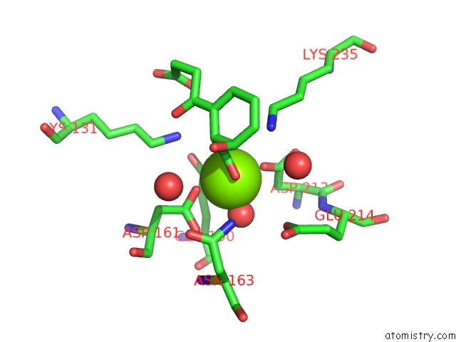
Mono view
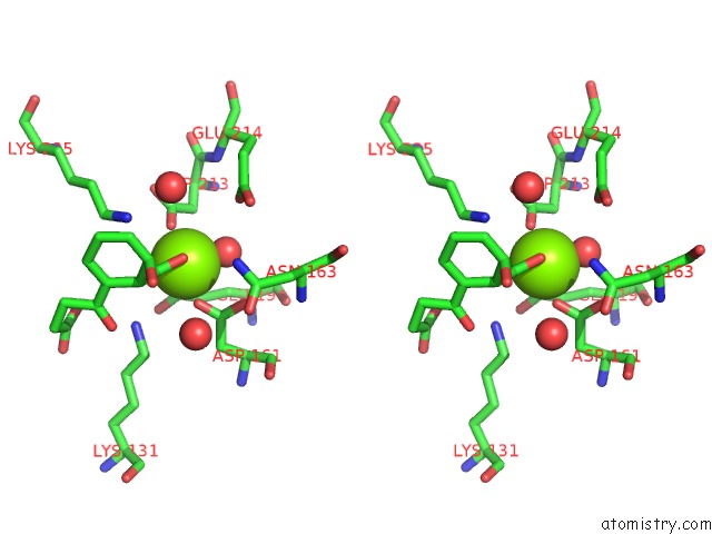
Stereo pair view
Mono view
Stereo pair view
A full contact list of Magnesium with other atoms in the Mg binding
site number 1 of Crystal Structure of the K133R Mutant of O-Succinylbenzoate Synthase (Osbs) From Escherichia Coli. Complex with Shchc within 5.0Å range:
|
Reference:
V.A.Klenchin,
E.A.Taylor Ringia,
J.A.Gerlt,
I.Rayment.
Evolution of Enzymatic Activity in the Enolase Superfamily: Structural and Mutagenic Studies of the Mechanism of the Reaction Catalyzed By O-Succinylbenzoate Synthase From Escherichia Coli Biochemistry V. 42 14427 2003.
ISSN: ISSN 0006-2960
PubMed: 14661953
DOI: 10.1021/BI035545V
Page generated: Tue Aug 13 12:04:56 2024
ISSN: ISSN 0006-2960
PubMed: 14661953
DOI: 10.1021/BI035545V
Last articles
Fe in 6ZKLFe in 6ZKI
Fe in 6ZKK
Fe in 6ZKJ
Fe in 6ZKH
Fe in 6ZKG
Fe in 6ZKF
Fe in 6ZKE
Fe in 6ZKD
Fe in 6ZKC