Magnesium »
PDB 1rtz-1s8f »
1s2o »
Magnesium in PDB 1s2o: X-Ray Structure of the Sucrose-Phosphatase (Spp) From Synechocystis Sp. PCC6803 at 1.40 A Resolution
Enzymatic activity of X-Ray Structure of the Sucrose-Phosphatase (Spp) From Synechocystis Sp. PCC6803 at 1.40 A Resolution
All present enzymatic activity of X-Ray Structure of the Sucrose-Phosphatase (Spp) From Synechocystis Sp. PCC6803 at 1.40 A Resolution:
3.1.3.24;
3.1.3.24;
Protein crystallography data
The structure of X-Ray Structure of the Sucrose-Phosphatase (Spp) From Synechocystis Sp. PCC6803 at 1.40 A Resolution, PDB code: 1s2o
was solved by
S.Fieulaine,
J.E.Lunn,
F.Borel,
J.L.Ferrer,
with X-Ray Crystallography technique. A brief refinement statistics is given in the table below:
| Resolution Low / High (Å) | 20.00 / 1.40 |
| Space group | P 1 21 1 |
| Cell size a, b, c (Å), α, β, γ (°) | 46.070, 51.760, 52.140, 90.00, 101.70, 90.00 |
| R / Rfree (%) | 18.2 / 20 |
Magnesium Binding Sites:
The binding sites of Magnesium atom in the X-Ray Structure of the Sucrose-Phosphatase (Spp) From Synechocystis Sp. PCC6803 at 1.40 A Resolution
(pdb code 1s2o). This binding sites where shown within
5.0 Angstroms radius around Magnesium atom.
In total only one binding site of Magnesium was determined in the X-Ray Structure of the Sucrose-Phosphatase (Spp) From Synechocystis Sp. PCC6803 at 1.40 A Resolution, PDB code: 1s2o:
In total only one binding site of Magnesium was determined in the X-Ray Structure of the Sucrose-Phosphatase (Spp) From Synechocystis Sp. PCC6803 at 1.40 A Resolution, PDB code: 1s2o:
Magnesium binding site 1 out of 1 in 1s2o
Go back to
Magnesium binding site 1 out
of 1 in the X-Ray Structure of the Sucrose-Phosphatase (Spp) From Synechocystis Sp. PCC6803 at 1.40 A Resolution
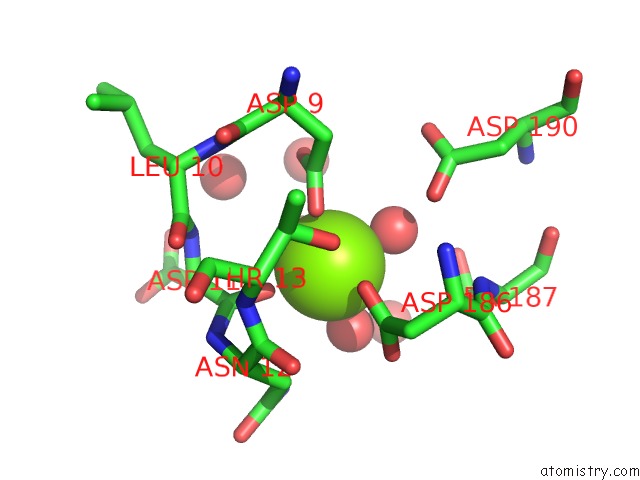
Mono view
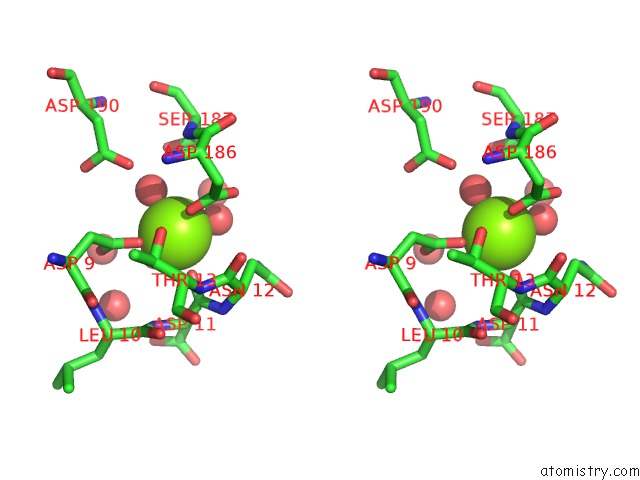
Stereo pair view
Mono view
Stereo pair view
A full contact list of Magnesium with other atoms in the Mg binding
site number 1 of X-Ray Structure of the Sucrose-Phosphatase (Spp) From Synechocystis Sp. PCC6803 at 1.40 A Resolution within 5.0Å range:
|
Reference:
S.Fieulaine,
J.E.Lunn,
F.Borel,
J.L.Ferrer.
The Structure of A Cyanobacterial Sucrose-Phosphatase Reveals the Sugar Tongs That Release Free Sucrose in the Cell. Plant Cell V. 17 2049 2005.
ISSN: ISSN 1040-4651
PubMed: 15937230
DOI: 10.1105/TPC.105.031229
Page generated: Sun Aug 10 04:02:54 2025
ISSN: ISSN 1040-4651
PubMed: 15937230
DOI: 10.1105/TPC.105.031229
Last articles
Mg in 428DMg in 412D
Mg in 421P
Mg in 3ZYY
Mg in 403D
Mg in 3ZYC
Mg in 3ZXW
Mg in 3ZXT
Mg in 3ZXD
Mg in 3ZXS