Magnesium »
PDB 1u3g-1uet »
1ue4 »
Magnesium in PDB 1ue4: Crystal Structure of D(Gcgaaagc)
Protein crystallography data
The structure of Crystal Structure of D(Gcgaaagc), PDB code: 1ue4
was solved by
T.Sunami,
J.Kondo,
I.Hirao,
K.Watanabe,
K.Miura,
A.Takenaka,
with X-Ray Crystallography technique. A brief refinement statistics is given in the table below:
| Resolution Low / High (Å) | 15.00 / 1.65 |
| Space group | I 4 2 2 |
| Cell size a, b, c (Å), α, β, γ (°) | 36.907, 36.907, 64.312, 90.00, 90.00, 90.00 |
| R / Rfree (%) | 20.9 / 23.6 |
Magnesium Binding Sites:
The binding sites of Magnesium atom in the Crystal Structure of D(Gcgaaagc)
(pdb code 1ue4). This binding sites where shown within
5.0 Angstroms radius around Magnesium atom.
In total only one binding site of Magnesium was determined in the Crystal Structure of D(Gcgaaagc), PDB code: 1ue4:
In total only one binding site of Magnesium was determined in the Crystal Structure of D(Gcgaaagc), PDB code: 1ue4:
Magnesium binding site 1 out of 1 in 1ue4
Go back to
Magnesium binding site 1 out
of 1 in the Crystal Structure of D(Gcgaaagc)
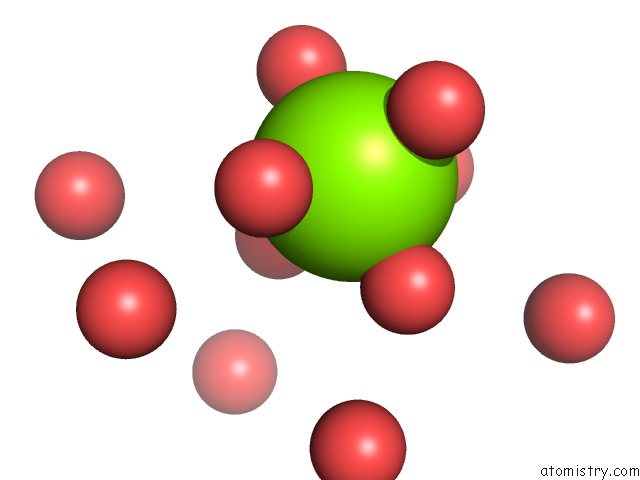
Mono view
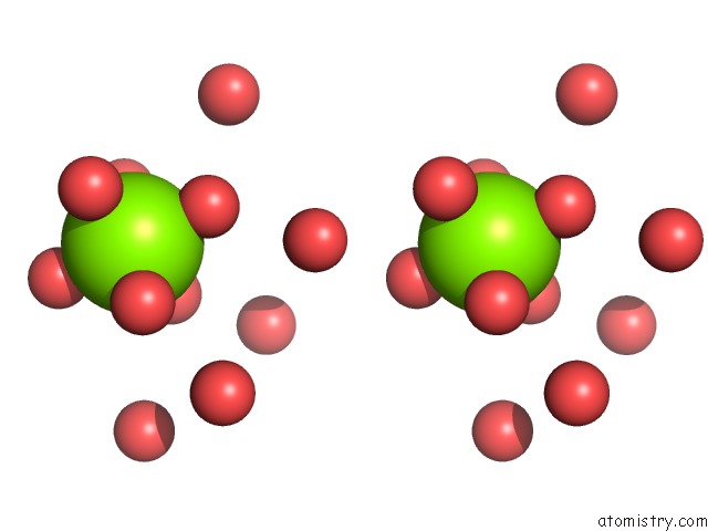
Stereo pair view
Mono view
Stereo pair view
A full contact list of Magnesium with other atoms in the Mg binding
site number 1 of Crystal Structure of D(Gcgaaagc) within 5.0Å range:
|
Reference:
T.Sunami,
J.Kondo,
I.Hirao,
K.Watanabe,
K.Miura,
A.Takenaka.
Structures of D(Gcgaagc) and D(Gcgaaagc) (Tetragonal Form): A Switching of Partners of the Sheared G.A Pairs to Form A Functional G.Axa.G Crossing. Acta Crystallogr.,Sect.D V. 60 422 2004.
ISSN: ISSN 0907-4449
PubMed: 14993665
DOI: 10.1107/S0907444903028415
Page generated: Sun Aug 10 05:05:51 2025
ISSN: ISSN 0907-4449
PubMed: 14993665
DOI: 10.1107/S0907444903028415
Last articles
Mg in 6L5NMg in 6L59
Mg in 6L5L
Mg in 6L4N
Mg in 6L57
Mg in 6L54
Mg in 6L3R
Mg in 6L42
Mg in 6L3E
Mg in 6L3G