Magnesium »
PDB 1v5g-1vq4 »
1v8s »
Magnesium in PDB 1v8s: Crystal Structure Analusis of the Adp-Ribose Pyrophosphatase Complexed with Amp and Mg
Enzymatic activity of Crystal Structure Analusis of the Adp-Ribose Pyrophosphatase Complexed with Amp and Mg
All present enzymatic activity of Crystal Structure Analusis of the Adp-Ribose Pyrophosphatase Complexed with Amp and Mg:
3.6.1.13;
3.6.1.13;
Protein crystallography data
The structure of Crystal Structure Analusis of the Adp-Ribose Pyrophosphatase Complexed with Amp and Mg, PDB code: 1v8s
was solved by
S.Yoshiba,
T.Ooga,
N.Nakagawa,
T.Shibata,
Y.Inoue,
S.Yokoyama,
S.Kuramitsu,
R.Masui,
Riken Structural Genomics/Proteomics Initiative (Rsgi),
with X-Ray Crystallography technique. A brief refinement statistics is given in the table below:
| Resolution Low / High (Å) | 34.80 / 2.20 |
| Space group | P 32 2 1 |
| Cell size a, b, c (Å), α, β, γ (°) | 49.740, 49.740, 118.120, 90.00, 90.00, 120.00 |
| R / Rfree (%) | 19.9 / 24.8 |
Magnesium Binding Sites:
The binding sites of Magnesium atom in the Crystal Structure Analusis of the Adp-Ribose Pyrophosphatase Complexed with Amp and Mg
(pdb code 1v8s). This binding sites where shown within
5.0 Angstroms radius around Magnesium atom.
In total only one binding site of Magnesium was determined in the Crystal Structure Analusis of the Adp-Ribose Pyrophosphatase Complexed with Amp and Mg, PDB code: 1v8s:
In total only one binding site of Magnesium was determined in the Crystal Structure Analusis of the Adp-Ribose Pyrophosphatase Complexed with Amp and Mg, PDB code: 1v8s:
Magnesium binding site 1 out of 1 in 1v8s
Go back to
Magnesium binding site 1 out
of 1 in the Crystal Structure Analusis of the Adp-Ribose Pyrophosphatase Complexed with Amp and Mg
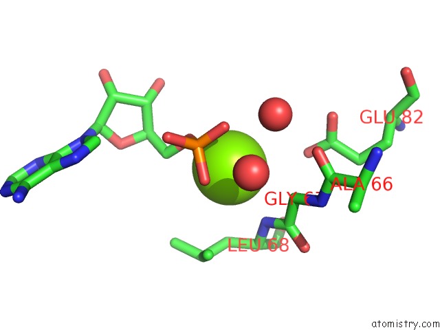
Mono view
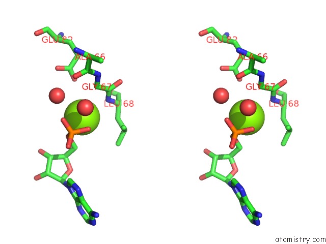
Stereo pair view
Mono view
Stereo pair view
A full contact list of Magnesium with other atoms in the Mg binding
site number 1 of Crystal Structure Analusis of the Adp-Ribose Pyrophosphatase Complexed with Amp and Mg within 5.0Å range:
|
Reference:
S.Yoshiba,
T.Ooga,
N.Nakagawa,
T.Shibata,
Y.Inoue,
S.Yokoyama,
S.Kuramitsu,
R.Masui.
Structural Insights Into the Thermus Thermophilus Adp-Ribose Pyrophosphatase Mechanism Via Crystal Structures with the Bound Substrate and Metal J.Biol.Chem. V. 279 37163 2004.
ISSN: ISSN 0021-9258
PubMed: 15210687
DOI: 10.1074/JBC.M403817200
Page generated: Sun Aug 10 05:14:08 2025
ISSN: ISSN 0021-9258
PubMed: 15210687
DOI: 10.1074/JBC.M403817200
Last articles
Mn in 9LJUMn in 9LJW
Mn in 9LJS
Mn in 9LJR
Mn in 9LJT
Mn in 9LJV
Mg in 9UA2
Mg in 9R96
Mg in 9VM1
Mg in 9P01