Magnesium »
PDB 1v5g-1vq4 »
1vfn »
Magnesium in PDB 1vfn: Purine Nucleoside Phosphorylase
Enzymatic activity of Purine Nucleoside Phosphorylase
All present enzymatic activity of Purine Nucleoside Phosphorylase:
2.4.2.1;
2.4.2.1;
Protein crystallography data
The structure of Purine Nucleoside Phosphorylase, PDB code: 1vfn
was solved by
G.Koellner,
A.Bzowska,
with X-Ray Crystallography technique. A brief refinement statistics is given in the table below:
| Resolution Low / High (Å) | 20.00 / 2.15 |
| Space group | P 21 3 |
| Cell size a, b, c (Å), α, β, γ (°) | 94.110, 94.110, 94.110, 90.00, 90.00, 90.00 |
| R / Rfree (%) | n/a / n/a |
Other elements in 1vfn:
The structure of Purine Nucleoside Phosphorylase also contains other interesting chemical elements:
| Zinc | (Zn) | 1 atom |
Magnesium Binding Sites:
The binding sites of Magnesium atom in the Purine Nucleoside Phosphorylase
(pdb code 1vfn). This binding sites where shown within
5.0 Angstroms radius around Magnesium atom.
In total only one binding site of Magnesium was determined in the Purine Nucleoside Phosphorylase, PDB code: 1vfn:
In total only one binding site of Magnesium was determined in the Purine Nucleoside Phosphorylase, PDB code: 1vfn:
Magnesium binding site 1 out of 1 in 1vfn
Go back to
Magnesium binding site 1 out
of 1 in the Purine Nucleoside Phosphorylase
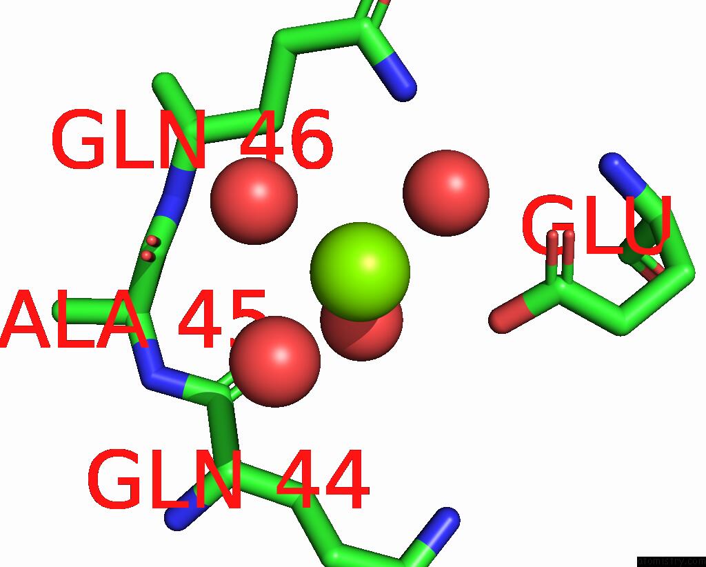
Mono view
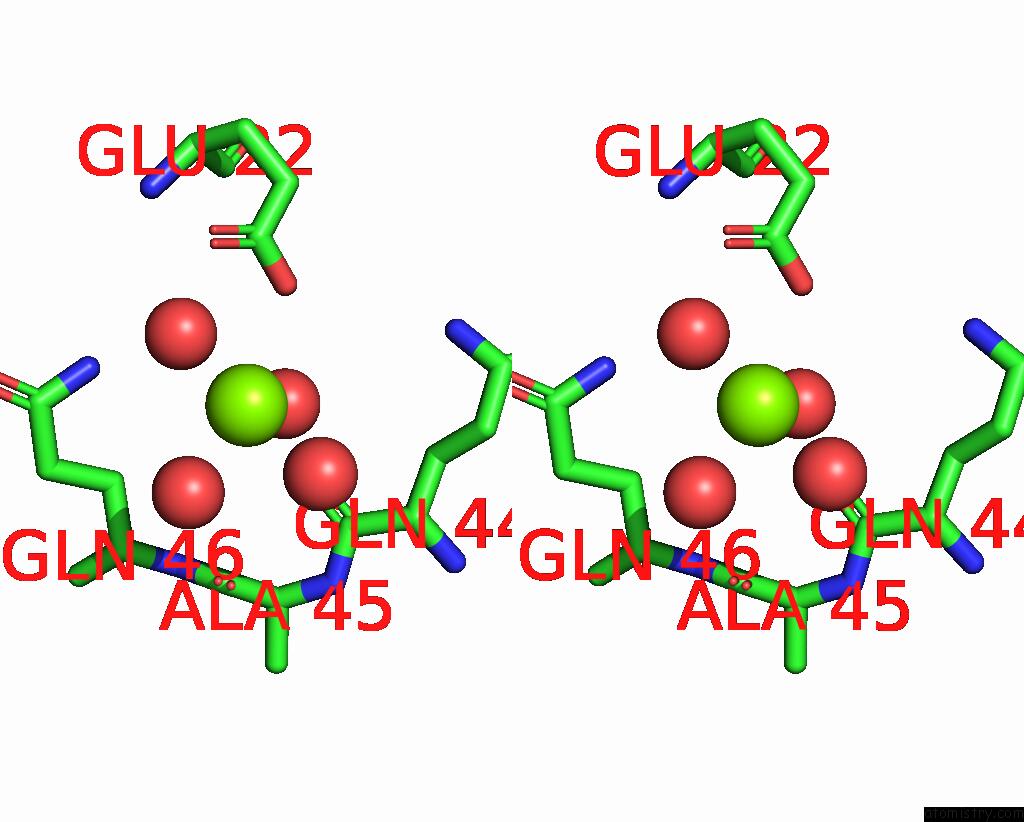
Stereo pair view
Mono view
Stereo pair view
A full contact list of Magnesium with other atoms in the Mg binding
site number 1 of Purine Nucleoside Phosphorylase within 5.0Å range:
|
Reference:
G.Koellner,
M.Luic,
D.Shugar,
W.Saenger,
A.Bzowska.
Crystal Structure of Calf Spleen Purine Nucleoside Phosphorylase in A Complex with Hypoxanthine at 2.15 A Resolution. J.Mol.Biol. V. 265 202 1997.
ISSN: ISSN 0022-2836
PubMed: 9020983
DOI: 10.1006/JMBI.1996.0730
Page generated: Sun Aug 10 05:15:29 2025
ISSN: ISSN 0022-2836
PubMed: 9020983
DOI: 10.1006/JMBI.1996.0730
Last articles
Fe in 9VR0Fe in 9UD8
Fe in 9QDT
Fe in 9S2T
Fe in 9JQA
Fe in 9IYV
Fe in 9J47
Fe in 9JQM
Fe in 9J2L
Fe in 9J1Q