Magnesium »
PDB 1vq5-1w55 »
1vr0 »
Magnesium in PDB 1vr0: Crystal Structure of Putative 2-Phosphosulfolactate Phosphatase (15026306) From Clostridium Acetobutylicum at 2.6 A Resolution
Enzymatic activity of Crystal Structure of Putative 2-Phosphosulfolactate Phosphatase (15026306) From Clostridium Acetobutylicum at 2.6 A Resolution
All present enzymatic activity of Crystal Structure of Putative 2-Phosphosulfolactate Phosphatase (15026306) From Clostridium Acetobutylicum at 2.6 A Resolution:
3.1.3.71;
3.1.3.71;
Protein crystallography data
The structure of Crystal Structure of Putative 2-Phosphosulfolactate Phosphatase (15026306) From Clostridium Acetobutylicum at 2.6 A Resolution, PDB code: 1vr0
was solved by
Joint Center For Structural Genomics (Jcsg),
with X-Ray Crystallography technique. A brief refinement statistics is given in the table below:
| Resolution Low / High (Å) | 38.70 / 2.49 |
| Space group | C 2 2 21 |
| Cell size a, b, c (Å), α, β, γ (°) | 46.686, 69.193, 453.518, 90.00, 90.00, 90.00 |
| R / Rfree (%) | 19 / 22.9 |
Magnesium Binding Sites:
The binding sites of Magnesium atom in the Crystal Structure of Putative 2-Phosphosulfolactate Phosphatase (15026306) From Clostridium Acetobutylicum at 2.6 A Resolution
(pdb code 1vr0). This binding sites where shown within
5.0 Angstroms radius around Magnesium atom.
In total 2 binding sites of Magnesium where determined in the Crystal Structure of Putative 2-Phosphosulfolactate Phosphatase (15026306) From Clostridium Acetobutylicum at 2.6 A Resolution, PDB code: 1vr0:
Jump to Magnesium binding site number: 1; 2;
In total 2 binding sites of Magnesium where determined in the Crystal Structure of Putative 2-Phosphosulfolactate Phosphatase (15026306) From Clostridium Acetobutylicum at 2.6 A Resolution, PDB code: 1vr0:
Jump to Magnesium binding site number: 1; 2;
Magnesium binding site 1 out of 2 in 1vr0
Go back to
Magnesium binding site 1 out
of 2 in the Crystal Structure of Putative 2-Phosphosulfolactate Phosphatase (15026306) From Clostridium Acetobutylicum at 2.6 A Resolution
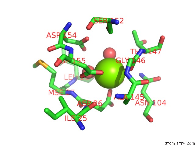
Mono view
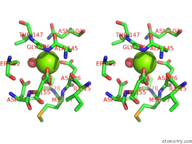
Stereo pair view
Mono view
Stereo pair view
A full contact list of Magnesium with other atoms in the Mg binding
site number 1 of Crystal Structure of Putative 2-Phosphosulfolactate Phosphatase (15026306) From Clostridium Acetobutylicum at 2.6 A Resolution within 5.0Å range:
|
Magnesium binding site 2 out of 2 in 1vr0
Go back to
Magnesium binding site 2 out
of 2 in the Crystal Structure of Putative 2-Phosphosulfolactate Phosphatase (15026306) From Clostridium Acetobutylicum at 2.6 A Resolution
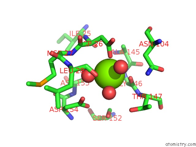
Mono view
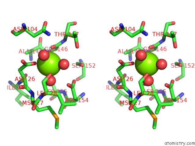
Stereo pair view
Mono view
Stereo pair view
A full contact list of Magnesium with other atoms in the Mg binding
site number 2 of Crystal Structure of Putative 2-Phosphosulfolactate Phosphatase (15026306) From Clostridium Acetobutylicum at 2.6 A Resolution within 5.0Å range:
|
Reference:
M.Didonato,
S.S.Krishna,
R.Schwarzenbacher,
D.Mcmullan,
S.Agarwalla,
S.M.Brittain,
M.D.Miller,
P.Abdubek,
E.Ambing,
H.L.Axelrod,
J.M.Canaves,
H.J.Chiu,
A.M.Deacon,
L.Duan,
M.A.Elsliger,
A.Godzik,
S.K.Grzechnik,
J.Hale,
E.Hampton,
J.Haugen,
L.Jaroszewski,
K.K.Jin,
H.E.Klock,
M.W.Knuth,
E.Koesema,
A.Kreusch,
P.Kuhn,
S.A.Lesley,
I.Levin,
A.T.Morse,
E.Nigoghossian,
L.Okach,
S.Oommachen,
J.Paulsen,
K.Quijano,
R.Reyes,
C.L.Rife,
G.Spraggon,
R.C.Stevens,
H.Van Den Bedem,
A.White,
G.Wolf,
Q.Xu,
K.O.Hodgson,
J.Wooley,
I.A.Wilson.
Crystal Structure of 2-Phosphosulfolactate Phosphatase (Comb) From Clostridium Acetobutylicum at 2.6 A Resolution Reveals A New Fold with A Novel Active Site. Proteins V. 65 771 2006.
ISSN: ISSN 0887-3585
PubMed: 16927339
DOI: 10.1002/PROT.20978
Page generated: Sun Aug 10 06:16:10 2025
ISSN: ISSN 0887-3585
PubMed: 16927339
DOI: 10.1002/PROT.20978
Last articles
Mg in 4DUYMg in 4DR7
Mg in 4DR6
Mg in 4DR5
Mg in 4DUX
Mg in 4DUW
Mg in 4DUV
Mg in 4DUO
Mg in 4DUG
Mg in 4DTY