Magnesium »
PDB 1w56-1wdt »
1w6t »
Magnesium in PDB 1w6t: Crystal Structure of Octameric Enolase From Streptococcus Pneumoniae
Enzymatic activity of Crystal Structure of Octameric Enolase From Streptococcus Pneumoniae
All present enzymatic activity of Crystal Structure of Octameric Enolase From Streptococcus Pneumoniae:
4.2.1.11;
4.2.1.11;
Protein crystallography data
The structure of Crystal Structure of Octameric Enolase From Streptococcus Pneumoniae, PDB code: 1w6t
was solved by
S.Ehinger,
W.-D.Schubert,
S.Bergmann,
S.Hammerschmidt,
D.W.Heinz,
with X-Ray Crystallography technique. A brief refinement statistics is given in the table below:
| Resolution Low / High (Å) | 100.00 / 2.10 |
| Space group | I 4 |
| Cell size a, b, c (Å), α, β, γ (°) | 143.702, 143.702, 100.579, 90.00, 90.00, 90.00 |
| R / Rfree (%) | 14.8 / 18.7 |
Magnesium Binding Sites:
The binding sites of Magnesium atom in the Crystal Structure of Octameric Enolase From Streptococcus Pneumoniae
(pdb code 1w6t). This binding sites where shown within
5.0 Angstroms radius around Magnesium atom.
In total 2 binding sites of Magnesium where determined in the Crystal Structure of Octameric Enolase From Streptococcus Pneumoniae, PDB code: 1w6t:
Jump to Magnesium binding site number: 1; 2;
In total 2 binding sites of Magnesium where determined in the Crystal Structure of Octameric Enolase From Streptococcus Pneumoniae, PDB code: 1w6t:
Jump to Magnesium binding site number: 1; 2;
Magnesium binding site 1 out of 2 in 1w6t
Go back to
Magnesium binding site 1 out
of 2 in the Crystal Structure of Octameric Enolase From Streptococcus Pneumoniae
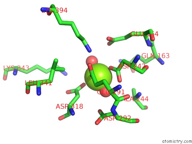
Mono view
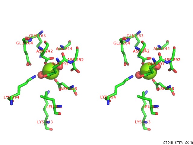
Stereo pair view
Mono view
Stereo pair view
A full contact list of Magnesium with other atoms in the Mg binding
site number 1 of Crystal Structure of Octameric Enolase From Streptococcus Pneumoniae within 5.0Å range:
|
Magnesium binding site 2 out of 2 in 1w6t
Go back to
Magnesium binding site 2 out
of 2 in the Crystal Structure of Octameric Enolase From Streptococcus Pneumoniae
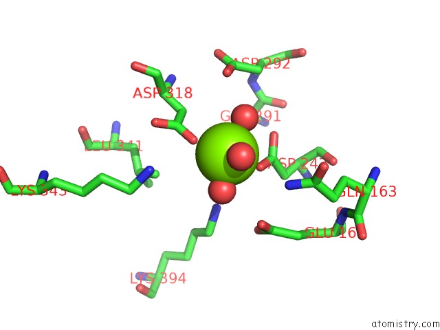
Mono view

Stereo pair view
Mono view
Stereo pair view
A full contact list of Magnesium with other atoms in the Mg binding
site number 2 of Crystal Structure of Octameric Enolase From Streptococcus Pneumoniae within 5.0Å range:
|
Reference:
S.Ehinger,
W.-D.Schubert,
S.Bergmann,
S.Hammerschmidt,
D.W.Heinz.
Plasmin(Ogen)-Binding Alpha-Enolase From Streptococcus Pneumoniae: Crystal Structure and Evaluation of Plasmin(Ogen)-Binding Sites J.Mol.Biol. V. 343 997 2004.
ISSN: ISSN 0022-2836
PubMed: 15476816
DOI: 10.1016/J.JMB.2004.08.088
Page generated: Tue Aug 13 17:08:55 2024
ISSN: ISSN 0022-2836
PubMed: 15476816
DOI: 10.1016/J.JMB.2004.08.088
Last articles
Fe in 2YXOFe in 2YRS
Fe in 2YXC
Fe in 2YNM
Fe in 2YVJ
Fe in 2YP1
Fe in 2YU2
Fe in 2YU1
Fe in 2YQB
Fe in 2YOO