Magnesium »
PDB 1xfy-1xon »
1xmx »
Magnesium in PDB 1xmx: Crystal Structure of Protein VC1899 From Vibrio Cholerae
Protein crystallography data
The structure of Crystal Structure of Protein VC1899 From Vibrio Cholerae, PDB code: 1xmx
was solved by
C.Chang,
A.Joachimiak,
R.Wu,
Midwest Center For Structural Genomics(Mcsg),
with X-Ray Crystallography technique. A brief refinement statistics is given in the table below:
| Resolution Low / High (Å) | 50.00 / 2.10 |
| Space group | P 21 21 2 |
| Cell size a, b, c (Å), α, β, γ (°) | 66.549, 71.439, 90.096, 90.00, 90.00, 90.00 |
| R / Rfree (%) | 16.8 / 25.4 |
Magnesium Binding Sites:
The binding sites of Magnesium atom in the Crystal Structure of Protein VC1899 From Vibrio Cholerae
(pdb code 1xmx). This binding sites where shown within
5.0 Angstroms radius around Magnesium atom.
In total only one binding site of Magnesium was determined in the Crystal Structure of Protein VC1899 From Vibrio Cholerae, PDB code: 1xmx:
In total only one binding site of Magnesium was determined in the Crystal Structure of Protein VC1899 From Vibrio Cholerae, PDB code: 1xmx:
Magnesium binding site 1 out of 1 in 1xmx
Go back to
Magnesium binding site 1 out
of 1 in the Crystal Structure of Protein VC1899 From Vibrio Cholerae
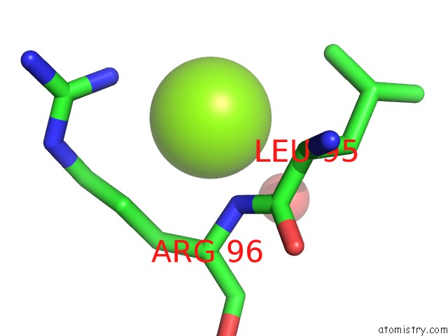
Mono view
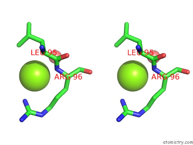
Stereo pair view
Mono view
Stereo pair view
A full contact list of Magnesium with other atoms in the Mg binding
site number 1 of Crystal Structure of Protein VC1899 From Vibrio Cholerae within 5.0Å range:
|
Reference:
C.Chang,
A.Joachimiak,
R.Wu,
S.Moy,
Midwest Center For Structural Genomics (Mcsg).
Crystal Structure of Protein VC1899 From Vibrio Cholerae To Be Published.
Page generated: Tue Aug 13 17:45:21 2024
Last articles
Ca in 3KCPCa in 3K9X
Ca in 3KCG
Ca in 3KAA
Ca in 3K9L
Ca in 3K9N
Ca in 3K71
Ca in 3K9J
Ca in 3K8Y
Ca in 3K8M