Magnesium »
PDB 1xoq-1xyc »
1xyc »
Magnesium in PDB 1xyc: X-Ray Crystallographic Structures of D-Xylose Isomerase-Substrate Complexes Position the Substrate and Provide Evidence For Metal Movement During Catalysis
Enzymatic activity of X-Ray Crystallographic Structures of D-Xylose Isomerase-Substrate Complexes Position the Substrate and Provide Evidence For Metal Movement During Catalysis
All present enzymatic activity of X-Ray Crystallographic Structures of D-Xylose Isomerase-Substrate Complexes Position the Substrate and Provide Evidence For Metal Movement During Catalysis:
5.3.1.5;
5.3.1.5;
Protein crystallography data
The structure of X-Ray Crystallographic Structures of D-Xylose Isomerase-Substrate Complexes Position the Substrate and Provide Evidence For Metal Movement During Catalysis, PDB code: 1xyc
was solved by
A.Lavie,
K.N.Allen,
G.A.Petsko,
D.Ringe,
with X-Ray Crystallography technique. A brief refinement statistics is given in the table below:
| Resolution Low / High (Å) | 10.00 / 2.19 |
| Space group | P 21 21 2 |
| Cell size a, b, c (Å), α, β, γ (°) | 87.700, 99.300, 94.300, 90.00, 90.00, 90.00 |
| R / Rfree (%) | 15.9 / n/a |
Magnesium Binding Sites:
The binding sites of Magnesium atom in the X-Ray Crystallographic Structures of D-Xylose Isomerase-Substrate Complexes Position the Substrate and Provide Evidence For Metal Movement During Catalysis
(pdb code 1xyc). This binding sites where shown within
5.0 Angstroms radius around Magnesium atom.
In total 6 binding sites of Magnesium where determined in the X-Ray Crystallographic Structures of D-Xylose Isomerase-Substrate Complexes Position the Substrate and Provide Evidence For Metal Movement During Catalysis, PDB code: 1xyc:
Jump to Magnesium binding site number: 1; 2; 3; 4; 5; 6;
In total 6 binding sites of Magnesium where determined in the X-Ray Crystallographic Structures of D-Xylose Isomerase-Substrate Complexes Position the Substrate and Provide Evidence For Metal Movement During Catalysis, PDB code: 1xyc:
Jump to Magnesium binding site number: 1; 2; 3; 4; 5; 6;
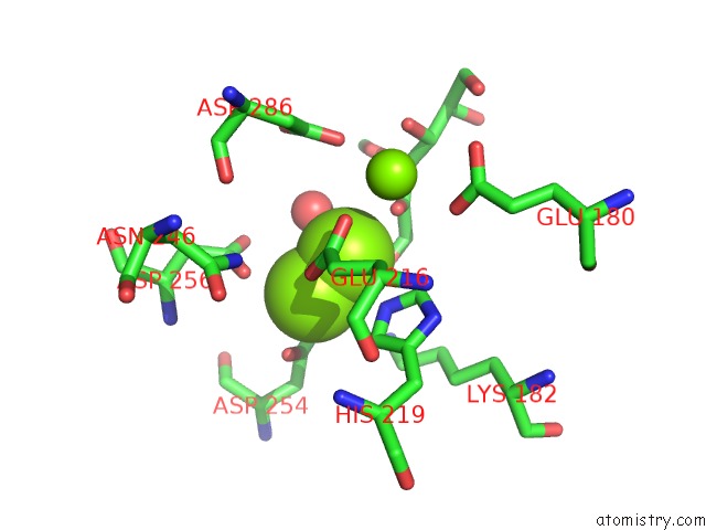
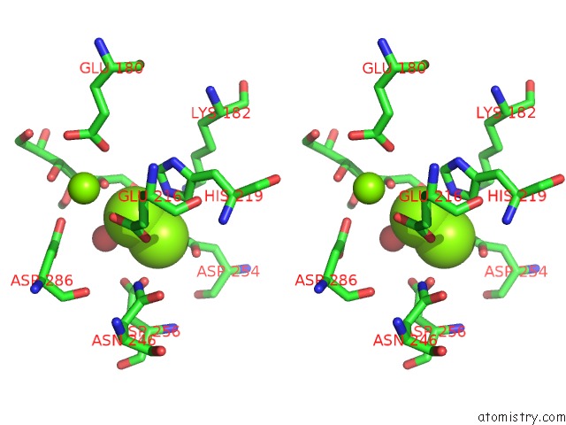
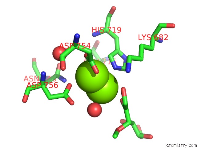
Magnesium binding site 1 out of 6 in 1xyc
Go back to
Magnesium binding site 1 out
of 6 in the X-Ray Crystallographic Structures of D-Xylose Isomerase-Substrate Complexes Position the Substrate and Provide Evidence For Metal Movement During Catalysis
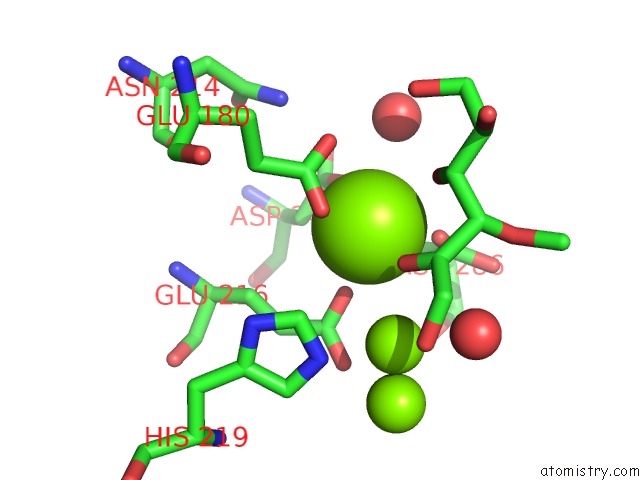
Mono view
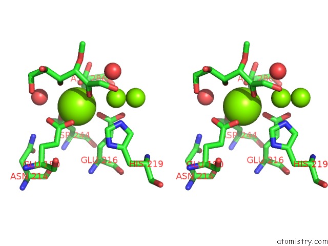
Stereo pair view
Mono view
Stereo pair view
A full contact list of Magnesium with other atoms in the Mg binding
site number 1 of X-Ray Crystallographic Structures of D-Xylose Isomerase-Substrate Complexes Position the Substrate and Provide Evidence For Metal Movement During Catalysis within 5.0Å range:
|
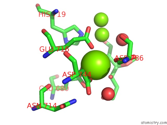
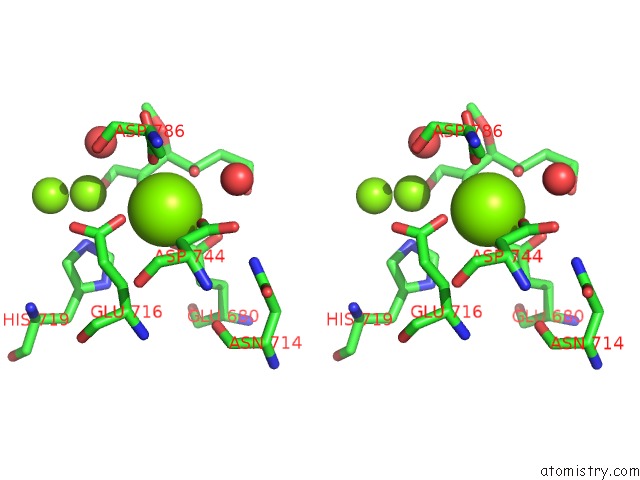
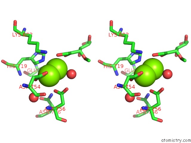
Magnesium binding site 2 out of 6 in 1xyc
Go back to
Magnesium binding site 2 out
of 6 in the X-Ray Crystallographic Structures of D-Xylose Isomerase-Substrate Complexes Position the Substrate and Provide Evidence For Metal Movement During Catalysis
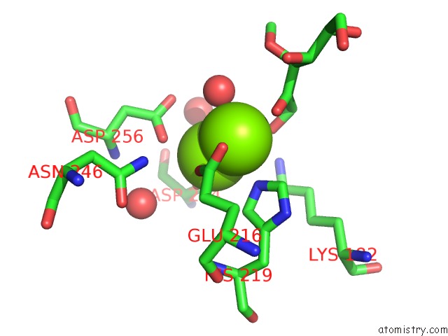
Mono view
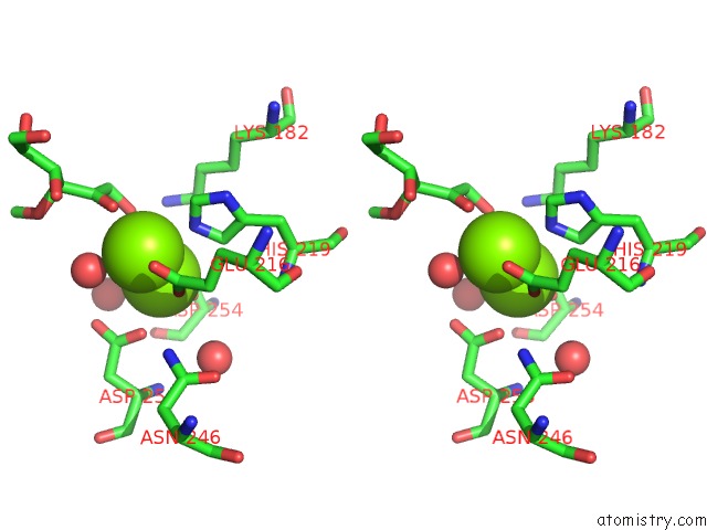
Stereo pair view
Mono view
Stereo pair view
A full contact list of Magnesium with other atoms in the Mg binding
site number 2 of X-Ray Crystallographic Structures of D-Xylose Isomerase-Substrate Complexes Position the Substrate and Provide Evidence For Metal Movement During Catalysis within 5.0Å range:
|
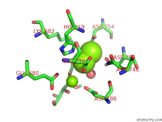
Magnesium binding site 3 out of 6 in 1xyc
Go back to
Magnesium binding site 3 out
of 6 in the X-Ray Crystallographic Structures of D-Xylose Isomerase-Substrate Complexes Position the Substrate and Provide Evidence For Metal Movement During Catalysis
Mono view
Stereo pair view
Mono view
Stereo pair view
A full contact list of Magnesium with other atoms in the Mg binding
site number 3 of X-Ray Crystallographic Structures of D-Xylose Isomerase-Substrate Complexes Position the Substrate and Provide Evidence For Metal Movement During Catalysis within 5.0Å range:
|
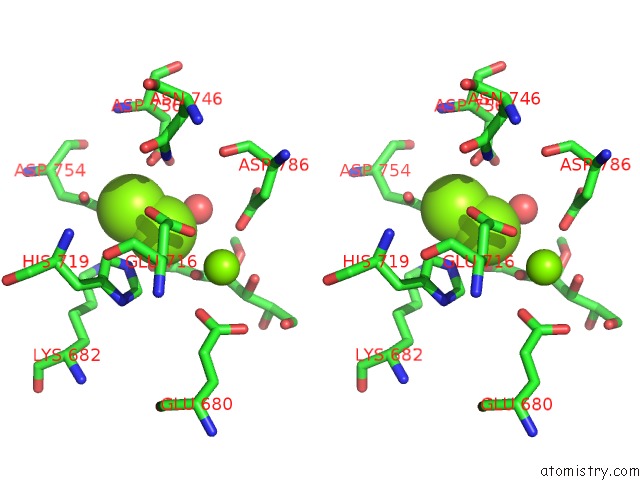
Magnesium binding site 4 out of 6 in 1xyc
Go back to
Magnesium binding site 4 out
of 6 in the X-Ray Crystallographic Structures of D-Xylose Isomerase-Substrate Complexes Position the Substrate and Provide Evidence For Metal Movement During Catalysis
Mono view
Stereo pair view
Mono view
Stereo pair view
A full contact list of Magnesium with other atoms in the Mg binding
site number 4 of X-Ray Crystallographic Structures of D-Xylose Isomerase-Substrate Complexes Position the Substrate and Provide Evidence For Metal Movement During Catalysis within 5.0Å range:
|
Magnesium binding site 5 out of 6 in 1xyc
Go back to
Magnesium binding site 5 out
of 6 in the X-Ray Crystallographic Structures of D-Xylose Isomerase-Substrate Complexes Position the Substrate and Provide Evidence For Metal Movement During Catalysis
Mono view
Stereo pair view
Mono view
Stereo pair view
A full contact list of Magnesium with other atoms in the Mg binding
site number 5 of X-Ray Crystallographic Structures of D-Xylose Isomerase-Substrate Complexes Position the Substrate and Provide Evidence For Metal Movement During Catalysis within 5.0Å range:
|
Magnesium binding site 6 out of 6 in 1xyc
Go back to
Magnesium binding site 6 out
of 6 in the X-Ray Crystallographic Structures of D-Xylose Isomerase-Substrate Complexes Position the Substrate and Provide Evidence For Metal Movement During Catalysis
Mono view
Stereo pair view
Mono view
Stereo pair view
A full contact list of Magnesium with other atoms in the Mg binding
site number 6 of X-Ray Crystallographic Structures of D-Xylose Isomerase-Substrate Complexes Position the Substrate and Provide Evidence For Metal Movement During Catalysis within 5.0Å range:
|
Reference:
A.Lavie,
K.N.Allen,
G.A.Petsko,
D.Ringe.
X-Ray Crystallographic Structures of D-Xylose Isomerase-Substrate Complexes Position the Substrate and Provide Evidence For Metal Movement During Catalysis. Biochemistry V. 33 5469 1994.
ISSN: ISSN 0006-2960
PubMed: 8180169
DOI: 10.1021/BI00184A016
Page generated: Sun Aug 10 07:38:17 2025
ISSN: ISSN 0006-2960
PubMed: 8180169
DOI: 10.1021/BI00184A016
Last articles
Mg in 5BTLMg in 5BTM
Mg in 5BTI
Mg in 5BTG
Mg in 5BTF
Mg in 5BTD
Mg in 5BTC
Mg in 5BTA
Mg in 5BON
Mg in 5BSU