Magnesium »
PDB 1ye8-1yqw »
1ykq »
Magnesium in PDB 1ykq: Crystal Structure of Diels-Alder Ribozyme
Protein crystallography data
The structure of Crystal Structure of Diels-Alder Ribozyme, PDB code: 1ykq
was solved by
A.Serganov,
S.Keiper,
L.Malinina,
V.Tereshko,
E.Skripkin,
C.Hobartner,
A.Polonskaia,
A.T.Phan,
R.Wombacher,
R.Micura,
Z.Dauter,
A.Jaschke,
D.J.Patel,
with X-Ray Crystallography technique. A brief refinement statistics is given in the table below:
| Resolution Low / High (Å) | 20.00 / 3.50 |
| Space group | P 1 21 1 |
| Cell size a, b, c (Å), α, β, γ (°) | 77.690, 44.240, 79.640, 90.00, 107.12, 90.00 |
| R / Rfree (%) | 28.6 / 32.4 |
Other elements in 1ykq:
The structure of Crystal Structure of Diels-Alder Ribozyme also contains other interesting chemical elements:
| Cadmium | (Cd) | 5 atoms |
Magnesium Binding Sites:
The binding sites of Magnesium atom in the Crystal Structure of Diels-Alder Ribozyme
(pdb code 1ykq). This binding sites where shown within
5.0 Angstroms radius around Magnesium atom.
In total 7 binding sites of Magnesium where determined in the Crystal Structure of Diels-Alder Ribozyme, PDB code: 1ykq:
Jump to Magnesium binding site number: 1; 2; 3; 4; 5; 6; 7;
In total 7 binding sites of Magnesium where determined in the Crystal Structure of Diels-Alder Ribozyme, PDB code: 1ykq:
Jump to Magnesium binding site number: 1; 2; 3; 4; 5; 6; 7;
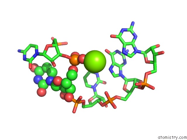
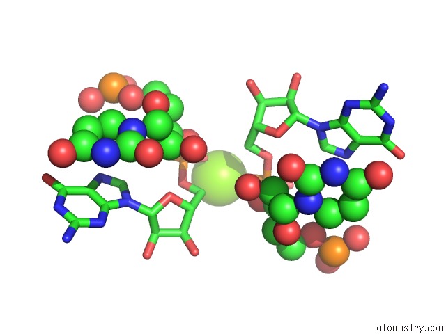
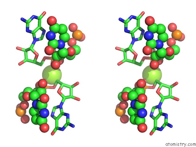
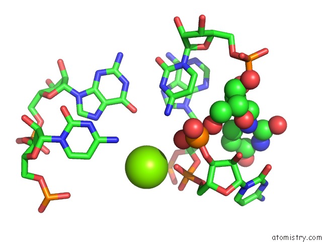
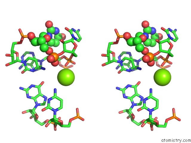
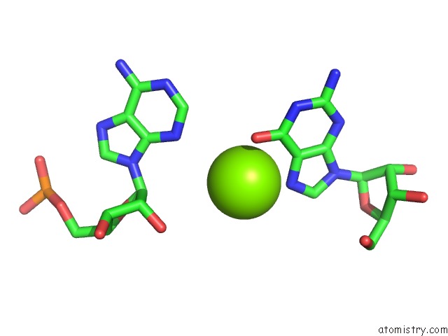
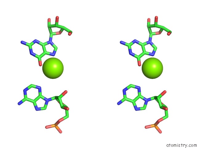
Magnesium binding site 1 out of 7 in 1ykq
Go back to
Magnesium binding site 1 out
of 7 in the Crystal Structure of Diels-Alder Ribozyme
Mono view
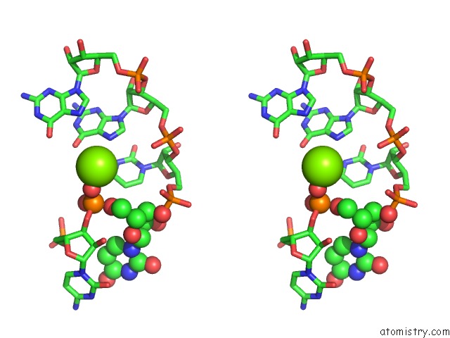
Stereo pair view
Mono view
Stereo pair view
A full contact list of Magnesium with other atoms in the Mg binding
site number 1 of Crystal Structure of Diels-Alder Ribozyme within 5.0Å range:
|
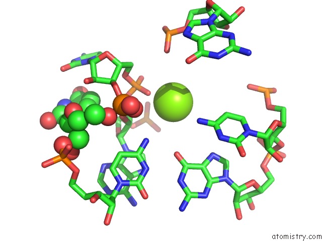
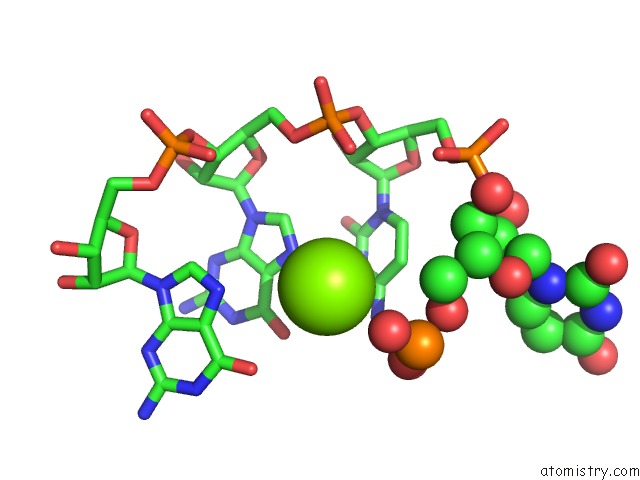
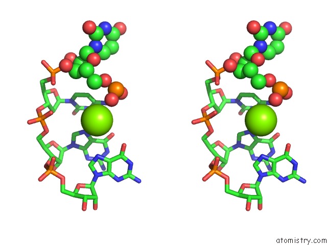
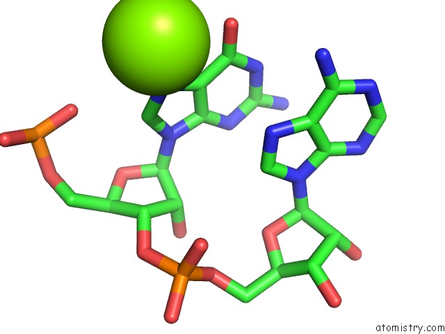
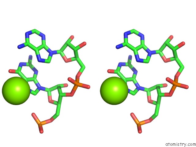
Magnesium binding site 2 out of 7 in 1ykq
Go back to
Magnesium binding site 2 out
of 7 in the Crystal Structure of Diels-Alder Ribozyme
Mono view
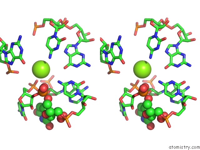
Stereo pair view
Mono view
Stereo pair view
A full contact list of Magnesium with other atoms in the Mg binding
site number 2 of Crystal Structure of Diels-Alder Ribozyme within 5.0Å range:
|
Magnesium binding site 3 out of 7 in 1ykq
Go back to
Magnesium binding site 3 out
of 7 in the Crystal Structure of Diels-Alder Ribozyme
Mono view
Stereo pair view
Mono view
Stereo pair view
A full contact list of Magnesium with other atoms in the Mg binding
site number 3 of Crystal Structure of Diels-Alder Ribozyme within 5.0Å range:
|
Magnesium binding site 4 out of 7 in 1ykq
Go back to
Magnesium binding site 4 out
of 7 in the Crystal Structure of Diels-Alder Ribozyme
Mono view
Stereo pair view
Mono view
Stereo pair view
A full contact list of Magnesium with other atoms in the Mg binding
site number 4 of Crystal Structure of Diels-Alder Ribozyme within 5.0Å range:
|
Magnesium binding site 5 out of 7 in 1ykq
Go back to
Magnesium binding site 5 out
of 7 in the Crystal Structure of Diels-Alder Ribozyme
Mono view
Stereo pair view
Mono view
Stereo pair view
A full contact list of Magnesium with other atoms in the Mg binding
site number 5 of Crystal Structure of Diels-Alder Ribozyme within 5.0Å range:
|
Magnesium binding site 6 out of 7 in 1ykq
Go back to
Magnesium binding site 6 out
of 7 in the Crystal Structure of Diels-Alder Ribozyme
Mono view
Stereo pair view
Mono view
Stereo pair view
A full contact list of Magnesium with other atoms in the Mg binding
site number 6 of Crystal Structure of Diels-Alder Ribozyme within 5.0Å range:
|
Magnesium binding site 7 out of 7 in 1ykq
Go back to
Magnesium binding site 7 out
of 7 in the Crystal Structure of Diels-Alder Ribozyme
Mono view
Stereo pair view
Mono view
Stereo pair view
A full contact list of Magnesium with other atoms in the Mg binding
site number 7 of Crystal Structure of Diels-Alder Ribozyme within 5.0Å range:
|
Reference:
A.Serganov,
S.Keiper,
L.Malinina,
V.Tereshko,
E.Skripkin,
C.Hobartner,
A.Polonskaia,
A.T.Phan,
R.Wombacher,
R.Micura,
Z.Dauter,
A.Jaschke,
D.J.Patel.
Structural Basis For Diels-Alder Ribozyme-Catalyzed Carbon-Carbon Bond Formation. Nat.Struct.Mol.Biol. V. 12 218 2005.
ISSN: ISSN 1545-9993
PubMed: 15723077
DOI: 10.1038/NSMB906
Page generated: Sun Aug 10 08:16:18 2025
ISSN: ISSN 1545-9993
PubMed: 15723077
DOI: 10.1038/NSMB906
Last articles
V in 1UZIV in 1YV3
V in 1VOM
V in 1VNI
V in 1VNH
V in 1VNG
V in 1VNF
V in 1VNE
V in 1VNC
V in 1VFZ