Magnesium »
PDB 1yqz-1yzt »
1yxo »
Magnesium in PDB 1yxo: Crystal Structure of Pyridoxal Phosphate Biosynthetic Protein Pdxa PA0593
Enzymatic activity of Crystal Structure of Pyridoxal Phosphate Biosynthetic Protein Pdxa PA0593
All present enzymatic activity of Crystal Structure of Pyridoxal Phosphate Biosynthetic Protein Pdxa PA0593:
1.1.1.262;
1.1.1.262;
Protein crystallography data
The structure of Crystal Structure of Pyridoxal Phosphate Biosynthetic Protein Pdxa PA0593, PDB code: 1yxo
was solved by
Y.Liu,
X.Xu,
A.Dong,
M.Kudritskam,
A.Savchenko,
E.F.Pai,
A.Joachimiak,
A.Edwards,
Midwest Center For Structural Genomics (Mcsg),
with X-Ray Crystallography technique. A brief refinement statistics is given in the table below:
| Resolution Low / High (Å) | 36.68 / 2.01 |
| Space group | P 21 21 21 |
| Cell size a, b, c (Å), α, β, γ (°) | 54.259, 59.307, 199.111, 90.00, 90.00, 90.00 |
| R / Rfree (%) | 19.8 / 24.3 |
Magnesium Binding Sites:
The binding sites of Magnesium atom in the Crystal Structure of Pyridoxal Phosphate Biosynthetic Protein Pdxa PA0593
(pdb code 1yxo). This binding sites where shown within
5.0 Angstroms radius around Magnesium atom.
In total 2 binding sites of Magnesium where determined in the Crystal Structure of Pyridoxal Phosphate Biosynthetic Protein Pdxa PA0593, PDB code: 1yxo:
Jump to Magnesium binding site number: 1; 2;
In total 2 binding sites of Magnesium where determined in the Crystal Structure of Pyridoxal Phosphate Biosynthetic Protein Pdxa PA0593, PDB code: 1yxo:
Jump to Magnesium binding site number: 1; 2;
Magnesium binding site 1 out of 2 in 1yxo
Go back to
Magnesium binding site 1 out
of 2 in the Crystal Structure of Pyridoxal Phosphate Biosynthetic Protein Pdxa PA0593
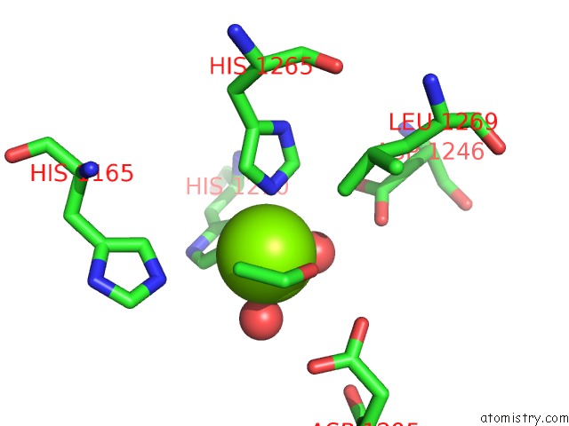
Mono view
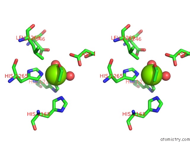
Stereo pair view
Mono view
Stereo pair view
A full contact list of Magnesium with other atoms in the Mg binding
site number 1 of Crystal Structure of Pyridoxal Phosphate Biosynthetic Protein Pdxa PA0593 within 5.0Å range:
|
Magnesium binding site 2 out of 2 in 1yxo
Go back to
Magnesium binding site 2 out
of 2 in the Crystal Structure of Pyridoxal Phosphate Biosynthetic Protein Pdxa PA0593
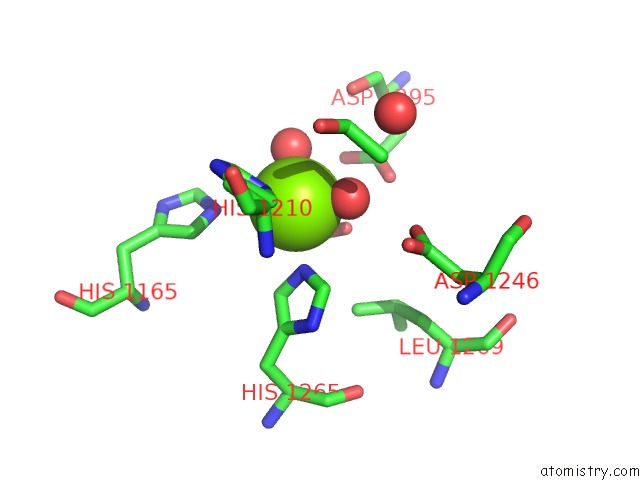
Mono view
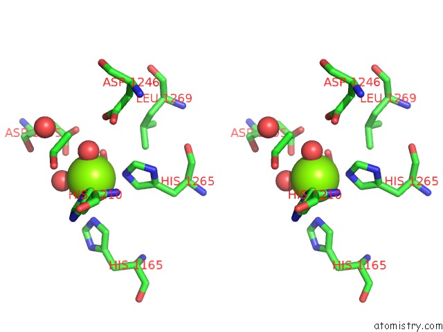
Stereo pair view
Mono view
Stereo pair view
A full contact list of Magnesium with other atoms in the Mg binding
site number 2 of Crystal Structure of Pyridoxal Phosphate Biosynthetic Protein Pdxa PA0593 within 5.0Å range:
|
Reference:
Y.Liu,
X.Xu,
A.Dong,
M.Kudritskam,
A.Savchenko,
E.F.Pai,
A.Edwards.
Crystal Structure of Pyridoxal Phosphate Biosynthetic Protein Pdxa PA0593 To Be Published.
Page generated: Tue Aug 13 19:55:56 2024
Last articles
Fe in 6N63Fe in 6N4M
Fe in 6N4L
Fe in 6N2N
Fe in 6N2O
Fe in 6N4J
Fe in 6N4K
Fe in 6N43
Fe in 6N21
Fe in 6N42