Magnesium »
PDB 1z6n-1zk2 »
1zbd »
Magnesium in PDB 1zbd: Structural Basis of Rab Effector Specificity: Crystal Structure of the Small G Protein RAB3A Complexed with the Effector Domain of Rabphilin-3A
Protein crystallography data
The structure of Structural Basis of Rab Effector Specificity: Crystal Structure of the Small G Protein RAB3A Complexed with the Effector Domain of Rabphilin-3A, PDB code: 1zbd
was solved by
C.Ostermeier,
A.T.Brunger,
with X-Ray Crystallography technique. A brief refinement statistics is given in the table below:
| Resolution Low / High (Å) | 90.00 / 2.60 |
| Space group | C 1 2 1 |
| Cell size a, b, c (Å), α, β, γ (°) | 89.315, 95.619, 47.715, 90.00, 94.49, 90.00 |
| R / Rfree (%) | 22.6 / 26.3 |
Other elements in 1zbd:
The structure of Structural Basis of Rab Effector Specificity: Crystal Structure of the Small G Protein RAB3A Complexed with the Effector Domain of Rabphilin-3A also contains other interesting chemical elements:
| Zinc | (Zn) | 2 atoms |
Magnesium Binding Sites:
The binding sites of Magnesium atom in the Structural Basis of Rab Effector Specificity: Crystal Structure of the Small G Protein RAB3A Complexed with the Effector Domain of Rabphilin-3A
(pdb code 1zbd). This binding sites where shown within
5.0 Angstroms radius around Magnesium atom.
In total only one binding site of Magnesium was determined in the Structural Basis of Rab Effector Specificity: Crystal Structure of the Small G Protein RAB3A Complexed with the Effector Domain of Rabphilin-3A, PDB code: 1zbd:
In total only one binding site of Magnesium was determined in the Structural Basis of Rab Effector Specificity: Crystal Structure of the Small G Protein RAB3A Complexed with the Effector Domain of Rabphilin-3A, PDB code: 1zbd:
Magnesium binding site 1 out of 1 in 1zbd
Go back to
Magnesium binding site 1 out
of 1 in the Structural Basis of Rab Effector Specificity: Crystal Structure of the Small G Protein RAB3A Complexed with the Effector Domain of Rabphilin-3A
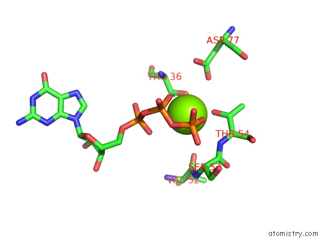
Mono view
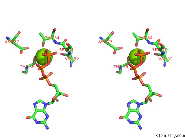
Stereo pair view
Mono view
Stereo pair view
A full contact list of Magnesium with other atoms in the Mg binding
site number 1 of Structural Basis of Rab Effector Specificity: Crystal Structure of the Small G Protein RAB3A Complexed with the Effector Domain of Rabphilin-3A within 5.0Å range:
|
Reference:
C.Ostermeier,
A.T.Brunger.
Structural Basis of Rab Effector Specificity: Crystal Structure of the Small G Protein RAB3A Complexed with the Effector Domain of Rabphilin-3A. Cell(Cambridge,Mass.) V. 96 363 1999.
ISSN: ISSN 0092-8674
PubMed: 10025402
DOI: 10.1016/S0092-8674(00)80549-8
Page generated: Sun Aug 10 08:49:02 2025
ISSN: ISSN 0092-8674
PubMed: 10025402
DOI: 10.1016/S0092-8674(00)80549-8
Last articles
Mn in 8ODOMn in 8ONX
Mn in 8OG4
Mn in 8ONE
Mn in 8OEO
Mn in 8OEH
Mn in 8OED
Mn in 8KFW
Mn in 8KFU
Mn in 8KFV