Magnesium »
PDB 2b2x-2bhc »
2bcj »
Magnesium in PDB 2bcj: Crystal Structure of G Protein-Coupled Receptor Kinase 2 in Complex with Galpha-Q and Gbetagamma Subunits
Enzymatic activity of Crystal Structure of G Protein-Coupled Receptor Kinase 2 in Complex with Galpha-Q and Gbetagamma Subunits
All present enzymatic activity of Crystal Structure of G Protein-Coupled Receptor Kinase 2 in Complex with Galpha-Q and Gbetagamma Subunits:
2.7.11.15;
2.7.11.15;
Protein crystallography data
The structure of Crystal Structure of G Protein-Coupled Receptor Kinase 2 in Complex with Galpha-Q and Gbetagamma Subunits, PDB code: 2bcj
was solved by
J.J.G.Tesmer,
with X-Ray Crystallography technique. A brief refinement statistics is given in the table below:
| Resolution Low / High (Å) | 46.78 / 3.06 |
| Space group | P 1 21 1 |
| Cell size a, b, c (Å), α, β, γ (°) | 64.918, 129.954, 122.764, 90.00, 95.81, 90.00 |
| R / Rfree (%) | 24.3 / 29.2 |
Other elements in 2bcj:
The structure of Crystal Structure of G Protein-Coupled Receptor Kinase 2 in Complex with Galpha-Q and Gbetagamma Subunits also contains other interesting chemical elements:
| Fluorine | (F) | 4 atoms |
| Aluminium | (Al) | 1 atom |
Magnesium Binding Sites:
The binding sites of Magnesium atom in the Crystal Structure of G Protein-Coupled Receptor Kinase 2 in Complex with Galpha-Q and Gbetagamma Subunits
(pdb code 2bcj). This binding sites where shown within
5.0 Angstroms radius around Magnesium atom.
In total only one binding site of Magnesium was determined in the Crystal Structure of G Protein-Coupled Receptor Kinase 2 in Complex with Galpha-Q and Gbetagamma Subunits, PDB code: 2bcj:
In total only one binding site of Magnesium was determined in the Crystal Structure of G Protein-Coupled Receptor Kinase 2 in Complex with Galpha-Q and Gbetagamma Subunits, PDB code: 2bcj:
Magnesium binding site 1 out of 1 in 2bcj
Go back to
Magnesium binding site 1 out
of 1 in the Crystal Structure of G Protein-Coupled Receptor Kinase 2 in Complex with Galpha-Q and Gbetagamma Subunits

Mono view
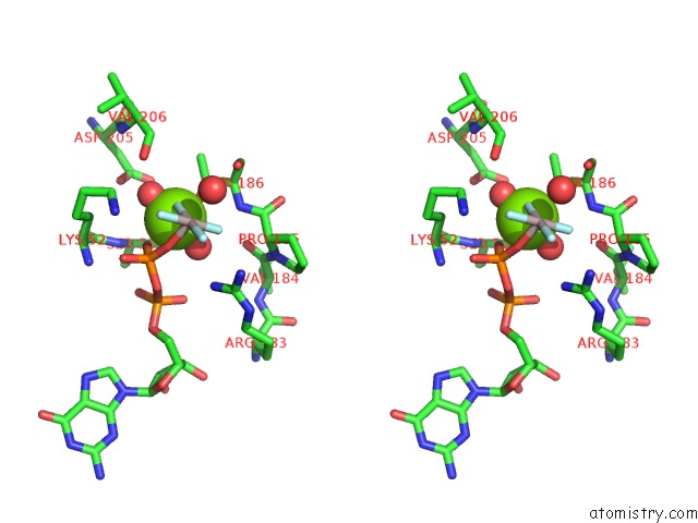
Stereo pair view
Mono view
Stereo pair view
A full contact list of Magnesium with other atoms in the Mg binding
site number 1 of Crystal Structure of G Protein-Coupled Receptor Kinase 2 in Complex with Galpha-Q and Gbetagamma Subunits within 5.0Å range:
|
Reference:
V.M.Tesmer,
T.Kawano,
A.Shankaranarayanan,
T.Kozasa,
J.J.G.Tesmer.
Snapshot of Activated G Proteins at the Membrane: the Gq-GRK2-G Complex Science V. 310 1686 2005.
ISSN: ISSN 0036-8075
PubMed: 16339447
DOI: 10.1126/SCIENCE.1118890
Page generated: Sun Aug 10 09:58:35 2025
ISSN: ISSN 0036-8075
PubMed: 16339447
DOI: 10.1126/SCIENCE.1118890
Last articles
Mg in 5GMKMg in 5GON
Mg in 5GPA
Mg in 5GP9
Mg in 5GOF
Mg in 5GMY
Mg in 5GOD
Mg in 5GKP
Mg in 5GKI
Mg in 5GKH