Magnesium »
PDB 2bt6-2c3y »
2c2j »
Magnesium in PDB 2c2j: Crystal Structure of the DPS92 From Deinococcus Radiodurans
Protein crystallography data
The structure of Crystal Structure of the DPS92 From Deinococcus Radiodurans, PDB code: 2c2j
was solved by
M.G.Cuypers,
C.V.Romao,
E.Mitchell,
S.Mcsweeney,
with X-Ray Crystallography technique. A brief refinement statistics is given in the table below:
| Resolution Low / High (Å) | 88.74 / 2.05 |
| Space group | P 2 3 |
| Cell size a, b, c (Å), α, β, γ (°) | 88.453, 88.453, 88.453, 90.00, 90.00, 90.00 |
| R / Rfree (%) | 17.9 / n/a |
Other elements in 2c2j:
The structure of Crystal Structure of the DPS92 From Deinococcus Radiodurans also contains other interesting chemical elements:
| Iron | (Fe) | 1 atom |
Magnesium Binding Sites:
The binding sites of Magnesium atom in the Crystal Structure of the DPS92 From Deinococcus Radiodurans
(pdb code 2c2j). This binding sites where shown within
5.0 Angstroms radius around Magnesium atom.
In total only one binding site of Magnesium was determined in the Crystal Structure of the DPS92 From Deinococcus Radiodurans, PDB code: 2c2j:
In total only one binding site of Magnesium was determined in the Crystal Structure of the DPS92 From Deinococcus Radiodurans, PDB code: 2c2j:
Magnesium binding site 1 out of 1 in 2c2j
Go back to
Magnesium binding site 1 out
of 1 in the Crystal Structure of the DPS92 From Deinococcus Radiodurans
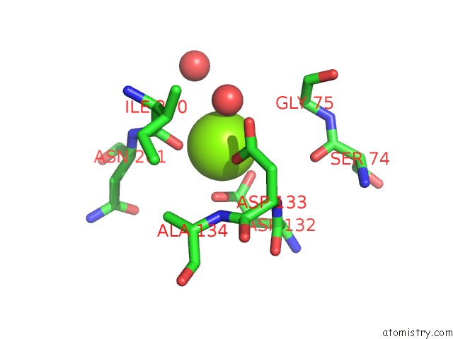
Mono view
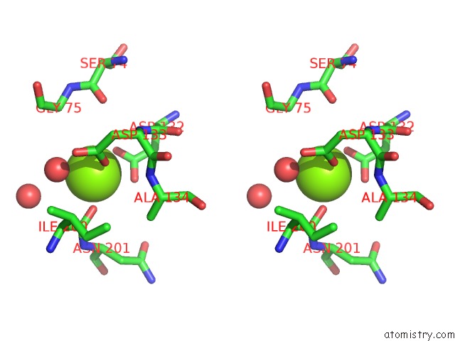
Stereo pair view
Mono view
Stereo pair view
A full contact list of Magnesium with other atoms in the Mg binding
site number 1 of Crystal Structure of the DPS92 From Deinococcus Radiodurans within 5.0Å range:
|
Reference:
M.G.Cuypers,
E.P.Mitchell,
C.V.Romao,
S.M.Mcsweeney.
The Crystal Structure of the DPS2 From Deinococcus Radiodurans Reveals An Unusual Pore Profile with A Non-Specific Metal Binding Site. J.Mol.Biol. V. 371 787 2007.
ISSN: ISSN 0022-2836
PubMed: 17583727
DOI: 10.1016/J.JMB.2006.11.032
Page generated: Tue Aug 13 22:08:50 2024
ISSN: ISSN 0022-2836
PubMed: 17583727
DOI: 10.1016/J.JMB.2006.11.032
Last articles
Fe in 2YXOFe in 2YRS
Fe in 2YXC
Fe in 2YNM
Fe in 2YVJ
Fe in 2YP1
Fe in 2YU2
Fe in 2YU1
Fe in 2YQB
Fe in 2YOO