Magnesium »
PDB 2c42-2cic »
2c6y »
Magnesium in PDB 2c6y: Crystal Structure of Interleukin Enhancer-Binding Factor 1 Bound to Dna
Protein crystallography data
The structure of Crystal Structure of Interleukin Enhancer-Binding Factor 1 Bound to Dna, PDB code: 2c6y
was solved by
K.-L.Tsai,
C.-Y.Huang,
C.-H.Chang,
Y.-J.Sun,
W.-J.Chuang,
C.-D.Hsiao,
with X-Ray Crystallography technique. A brief refinement statistics is given in the table below:
| Resolution Low / High (Å) | 29.44 / 2.4 |
| Space group | P 61 2 2 |
| Cell size a, b, c (Å), α, β, γ (°) | 58.736, 58.736, 324.923, 90.00, 90.00, 120.00 |
| R / Rfree (%) | 23.3 / 25.8 |
Magnesium Binding Sites:
The binding sites of Magnesium atom in the Crystal Structure of Interleukin Enhancer-Binding Factor 1 Bound to Dna
(pdb code 2c6y). This binding sites where shown within
5.0 Angstroms radius around Magnesium atom.
In total 2 binding sites of Magnesium where determined in the Crystal Structure of Interleukin Enhancer-Binding Factor 1 Bound to Dna, PDB code: 2c6y:
Jump to Magnesium binding site number: 1; 2;
In total 2 binding sites of Magnesium where determined in the Crystal Structure of Interleukin Enhancer-Binding Factor 1 Bound to Dna, PDB code: 2c6y:
Jump to Magnesium binding site number: 1; 2;
Magnesium binding site 1 out of 2 in 2c6y
Go back to
Magnesium binding site 1 out
of 2 in the Crystal Structure of Interleukin Enhancer-Binding Factor 1 Bound to Dna
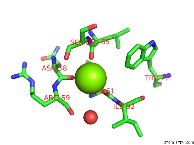
Mono view
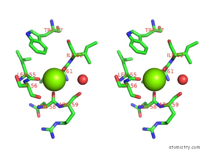
Stereo pair view
Mono view
Stereo pair view
A full contact list of Magnesium with other atoms in the Mg binding
site number 1 of Crystal Structure of Interleukin Enhancer-Binding Factor 1 Bound to Dna within 5.0Å range:
|
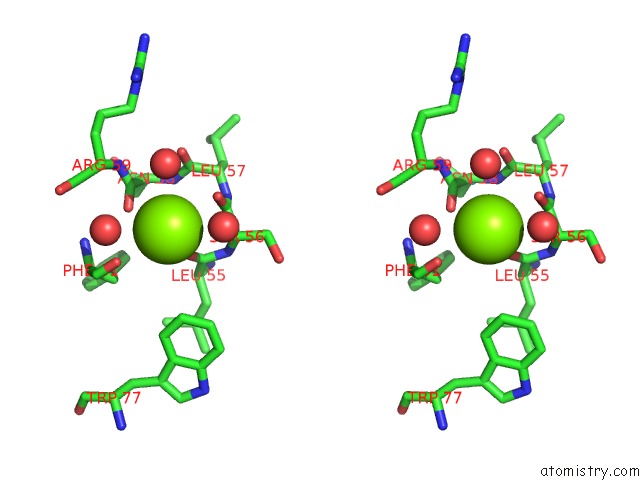
Magnesium binding site 2 out of 2 in 2c6y
Go back to
Magnesium binding site 2 out
of 2 in the Crystal Structure of Interleukin Enhancer-Binding Factor 1 Bound to Dna
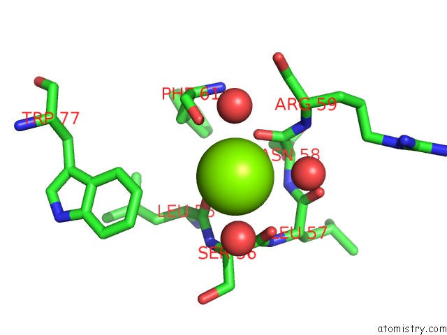
Mono view
Stereo pair view
Mono view
Stereo pair view
A full contact list of Magnesium with other atoms in the Mg binding
site number 2 of Crystal Structure of Interleukin Enhancer-Binding Factor 1 Bound to Dna within 5.0Å range:
|
Reference:
K.-L.Tsai,
C.-Y.Huang,
C.-H.Chang,
Y.-J.Sun,
W.-J.Chuang,
C.-D.Hsiao.
Crystal Structure of the Human FOXK1A-Dna Complex and Its Implications on the Diverse Binding Specificity of Winged Helix/Forkhead Proteins. J.Biol.Chem. V. 281 17400 2006.
ISSN: ISSN 0021-9258
PubMed: 16624804
DOI: 10.1074/JBC.M600478200
Page generated: Sun Aug 10 10:14:35 2025
ISSN: ISSN 0021-9258
PubMed: 16624804
DOI: 10.1074/JBC.M600478200
Last articles
Mg in 5GQVMg in 5GQM
Mg in 5GQL
Mg in 5GQ9
Mg in 5GPJ
Mg in 5GQJ
Mg in 5GL3
Mg in 5GQI
Mg in 5GMK
Mg in 5GON