Magnesium »
PDB 2c42-2cic »
2cg5 »
Magnesium in PDB 2cg5: Structure of Aminoadipate-Semialdehyde Dehydrogenase- Phosphopantetheinyl Transferase in Complex with Cytosolic Acyl Carrier Protein and Coenzyme A
Enzymatic activity of Structure of Aminoadipate-Semialdehyde Dehydrogenase- Phosphopantetheinyl Transferase in Complex with Cytosolic Acyl Carrier Protein and Coenzyme A
All present enzymatic activity of Structure of Aminoadipate-Semialdehyde Dehydrogenase- Phosphopantetheinyl Transferase in Complex with Cytosolic Acyl Carrier Protein and Coenzyme A:
1.2.1.31; 2.3.1.85;
1.2.1.31; 2.3.1.85;
Protein crystallography data
The structure of Structure of Aminoadipate-Semialdehyde Dehydrogenase- Phosphopantetheinyl Transferase in Complex with Cytosolic Acyl Carrier Protein and Coenzyme A, PDB code: 2cg5
was solved by
G.Bunkoczi,
A.Joshi,
E.Papagrigoriu,
C.Arrowsmith,
A.Edwards,
M.Sundstrom,
J.Weigelt,
F.Von Delft,
S.Smith,
U.Oppermann,
with X-Ray Crystallography technique. A brief refinement statistics is given in the table below:
| Resolution Low / High (Å) | 50.00 / 2.70 |
| Space group | P 32 2 1 |
| Cell size a, b, c (Å), α, β, γ (°) | 69.361, 69.361, 184.715, 90.00, 90.00, 120.00 |
| R / Rfree (%) | 19.2 / 24.7 |
Other elements in 2cg5:
The structure of Structure of Aminoadipate-Semialdehyde Dehydrogenase- Phosphopantetheinyl Transferase in Complex with Cytosolic Acyl Carrier Protein and Coenzyme A also contains other interesting chemical elements:
| Nickel | (Ni) | 1 atom |
Magnesium Binding Sites:
The binding sites of Magnesium atom in the Structure of Aminoadipate-Semialdehyde Dehydrogenase- Phosphopantetheinyl Transferase in Complex with Cytosolic Acyl Carrier Protein and Coenzyme A
(pdb code 2cg5). This binding sites where shown within
5.0 Angstroms radius around Magnesium atom.
In total 2 binding sites of Magnesium where determined in the Structure of Aminoadipate-Semialdehyde Dehydrogenase- Phosphopantetheinyl Transferase in Complex with Cytosolic Acyl Carrier Protein and Coenzyme A, PDB code: 2cg5:
Jump to Magnesium binding site number: 1; 2;
In total 2 binding sites of Magnesium where determined in the Structure of Aminoadipate-Semialdehyde Dehydrogenase- Phosphopantetheinyl Transferase in Complex with Cytosolic Acyl Carrier Protein and Coenzyme A, PDB code: 2cg5:
Jump to Magnesium binding site number: 1; 2;
Magnesium binding site 1 out of 2 in 2cg5
Go back to
Magnesium binding site 1 out
of 2 in the Structure of Aminoadipate-Semialdehyde Dehydrogenase- Phosphopantetheinyl Transferase in Complex with Cytosolic Acyl Carrier Protein and Coenzyme A
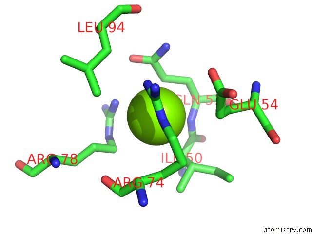
Mono view
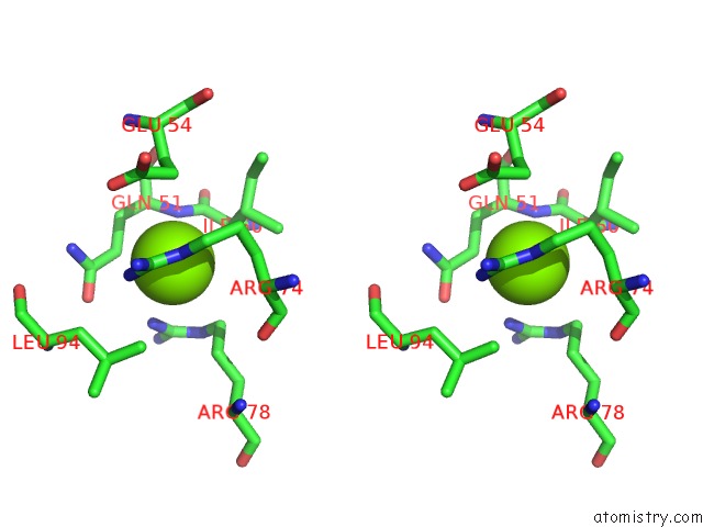
Stereo pair view
Mono view
Stereo pair view
A full contact list of Magnesium with other atoms in the Mg binding
site number 1 of Structure of Aminoadipate-Semialdehyde Dehydrogenase- Phosphopantetheinyl Transferase in Complex with Cytosolic Acyl Carrier Protein and Coenzyme A within 5.0Å range:
|
Magnesium binding site 2 out of 2 in 2cg5
Go back to
Magnesium binding site 2 out
of 2 in the Structure of Aminoadipate-Semialdehyde Dehydrogenase- Phosphopantetheinyl Transferase in Complex with Cytosolic Acyl Carrier Protein and Coenzyme A
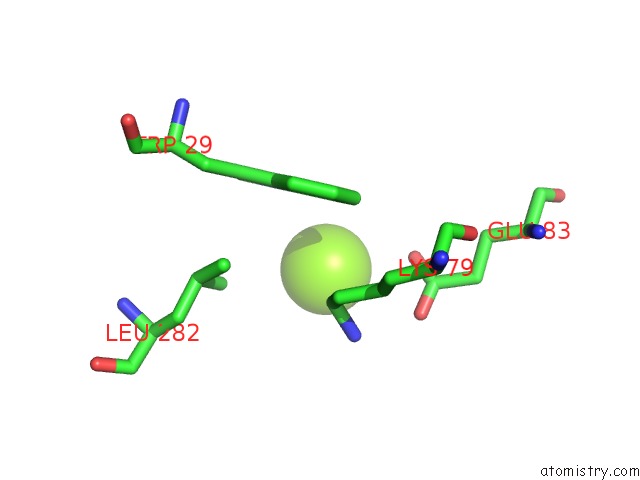
Mono view
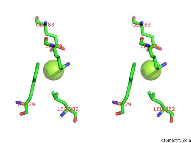
Stereo pair view
Mono view
Stereo pair view
A full contact list of Magnesium with other atoms in the Mg binding
site number 2 of Structure of Aminoadipate-Semialdehyde Dehydrogenase- Phosphopantetheinyl Transferase in Complex with Cytosolic Acyl Carrier Protein and Coenzyme A within 5.0Å range:
|
Reference:
G.Bunkoczi,
S.Pasta,
A.Joshi,
X.Wu,
K.L.Kavanagh,
S.Smith,
U.Oppermann.
Mechanism and Substrate Recognition of Human Holo Acp Synthase. Chem.Biol. V. 14 1243 2007.
ISSN: ISSN 1074-5521
PubMed: 18022563
DOI: 10.1016/J.CHEMBIOL.2007.10.013
Page generated: Tue Aug 13 22:15:44 2024
ISSN: ISSN 1074-5521
PubMed: 18022563
DOI: 10.1016/J.CHEMBIOL.2007.10.013
Last articles
Zn in 9MJ5Zn in 9HNW
Zn in 9G0L
Zn in 9FNE
Zn in 9DZN
Zn in 9E0I
Zn in 9D32
Zn in 9DAK
Zn in 8ZXC
Zn in 8ZUF