Magnesium »
PDB 2gqr-2haw »
2h5n »
Magnesium in PDB 2h5n: Crystal Structure of Protein of Unknown Function PG1108 From Porphyromonas Gingivalis W83
Protein crystallography data
The structure of Crystal Structure of Protein of Unknown Function PG1108 From Porphyromonas Gingivalis W83, PDB code: 2h5n
was solved by
B.Nocek,
L.Bigelow,
S.Moy,
A.Joachimiak,
Midwest Center For Structuralgenomics (Mcsg),
with X-Ray Crystallography technique. A brief refinement statistics is given in the table below:
| Resolution Low / High (Å) | 40.00 / 2.01 |
| Space group | C 2 2 21 |
| Cell size a, b, c (Å), α, β, γ (°) | 79.961, 84.826, 164.535, 90.00, 90.00, 90.00 |
| R / Rfree (%) | 18.8 / 24.4 |
Magnesium Binding Sites:
The binding sites of Magnesium atom in the Crystal Structure of Protein of Unknown Function PG1108 From Porphyromonas Gingivalis W83
(pdb code 2h5n). This binding sites where shown within
5.0 Angstroms radius around Magnesium atom.
In total 3 binding sites of Magnesium where determined in the Crystal Structure of Protein of Unknown Function PG1108 From Porphyromonas Gingivalis W83, PDB code: 2h5n:
Jump to Magnesium binding site number: 1; 2; 3;
In total 3 binding sites of Magnesium where determined in the Crystal Structure of Protein of Unknown Function PG1108 From Porphyromonas Gingivalis W83, PDB code: 2h5n:
Jump to Magnesium binding site number: 1; 2; 3;
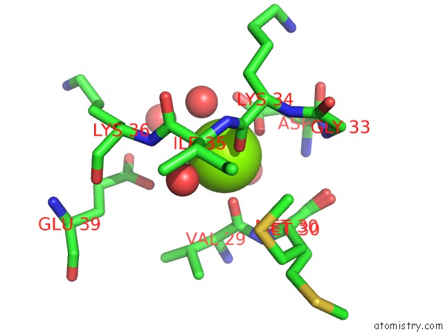
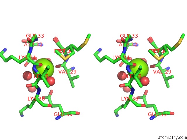
Magnesium binding site 1 out of 3 in 2h5n
Go back to
Magnesium binding site 1 out
of 3 in the Crystal Structure of Protein of Unknown Function PG1108 From Porphyromonas Gingivalis W83
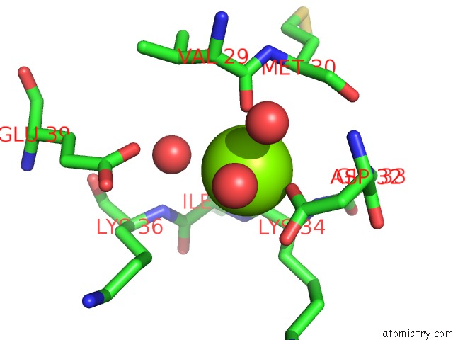
Mono view
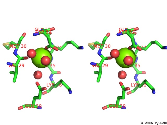
Stereo pair view
Mono view
Stereo pair view
A full contact list of Magnesium with other atoms in the Mg binding
site number 1 of Crystal Structure of Protein of Unknown Function PG1108 From Porphyromonas Gingivalis W83 within 5.0Å range:
|
Magnesium binding site 2 out of 3 in 2h5n
Go back to
Magnesium binding site 2 out
of 3 in the Crystal Structure of Protein of Unknown Function PG1108 From Porphyromonas Gingivalis W83
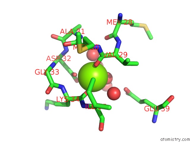
Mono view
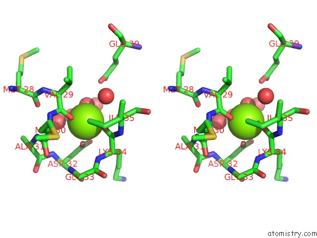
Stereo pair view
Mono view
Stereo pair view
A full contact list of Magnesium with other atoms in the Mg binding
site number 2 of Crystal Structure of Protein of Unknown Function PG1108 From Porphyromonas Gingivalis W83 within 5.0Å range:
|
Magnesium binding site 3 out of 3 in 2h5n
Go back to
Magnesium binding site 3 out
of 3 in the Crystal Structure of Protein of Unknown Function PG1108 From Porphyromonas Gingivalis W83
Mono view
Stereo pair view
Mono view
Stereo pair view
A full contact list of Magnesium with other atoms in the Mg binding
site number 3 of Crystal Structure of Protein of Unknown Function PG1108 From Porphyromonas Gingivalis W83 within 5.0Å range:
|
Reference:
B.Nocek,
L.Bigelow,
S.Moy,
A.Joachimiak.
Crystal Structure of Hypothetical Protein PG_1108 From Porphyromonas Gingivalis W83 To Be Published.
Page generated: Sun Aug 10 11:17:15 2025
Last articles
Mg in 4DUYMg in 4DR7
Mg in 4DR6
Mg in 4DR5
Mg in 4DUX
Mg in 4DUW
Mg in 4DUV
Mg in 4DUO
Mg in 4DUG
Mg in 4DTY