Magnesium »
PDB 2hb4-2hkj »
2hb5 »
Magnesium in PDB 2hb5: Crystal Structure of the Moloney Murine Leukemia Virus Rnase H Domain
Enzymatic activity of Crystal Structure of the Moloney Murine Leukemia Virus Rnase H Domain
All present enzymatic activity of Crystal Structure of the Moloney Murine Leukemia Virus Rnase H Domain:
3.1.26.4;
3.1.26.4;
Protein crystallography data
The structure of Crystal Structure of the Moloney Murine Leukemia Virus Rnase H Domain, PDB code: 2hb5
was solved by
D.Lim,
G.G.Gregorio,
C.Bingman,
E.Martinez-Hackert,
W.A.Hendrickson,
S.P.Goff,
with X-Ray Crystallography technique. A brief refinement statistics is given in the table below:
| Resolution Low / High (Å) | 21.66 / 1.59 |
| Space group | P 1 |
| Cell size a, b, c (Å), α, β, γ (°) | 32.326, 34.107, 34.802, 78.21, 69.85, 64.79 |
| R / Rfree (%) | 19.6 / 22.1 |
Magnesium Binding Sites:
The binding sites of Magnesium atom in the Crystal Structure of the Moloney Murine Leukemia Virus Rnase H Domain
(pdb code 2hb5). This binding sites where shown within
5.0 Angstroms radius around Magnesium atom.
In total only one binding site of Magnesium was determined in the Crystal Structure of the Moloney Murine Leukemia Virus Rnase H Domain, PDB code: 2hb5:
In total only one binding site of Magnesium was determined in the Crystal Structure of the Moloney Murine Leukemia Virus Rnase H Domain, PDB code: 2hb5:
Magnesium binding site 1 out of 1 in 2hb5
Go back to
Magnesium binding site 1 out
of 1 in the Crystal Structure of the Moloney Murine Leukemia Virus Rnase H Domain
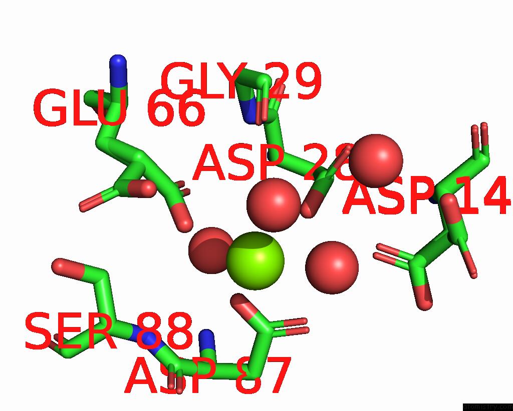
Mono view
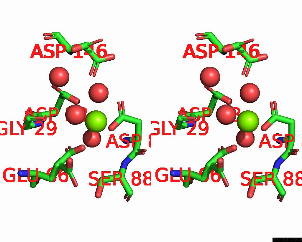
Stereo pair view
Mono view
Stereo pair view
A full contact list of Magnesium with other atoms in the Mg binding
site number 1 of Crystal Structure of the Moloney Murine Leukemia Virus Rnase H Domain within 5.0Å range:
|
Reference:
D.Lim,
G.G.Gregorio,
C.Bingman,
E.Martinez-Hackert,
W.A.Hendrickson,
S.P.Goff.
Crystal Structure of the Moloney Murine Leukemia Virus Rnase H Domain. J.Virol. V. 80 8379 2006.
ISSN: ISSN 0022-538X
PubMed: 16912289
DOI: 10.1128/JVI.00750-06
Page generated: Sun Aug 10 11:20:17 2025
ISSN: ISSN 0022-538X
PubMed: 16912289
DOI: 10.1128/JVI.00750-06
Last articles
Mg in 4DR2Mg in 4DR3
Mg in 4DR1
Mg in 4DPG
Mg in 4DQP
Mg in 4DQQ
Mg in 4DPM
Mg in 4DPV
Mg in 4DQI
Mg in 4DOB