Magnesium »
PDB 2o56-2oh6 »
2oek »
Magnesium in PDB 2oek: Crystal Structure of A Rubisco-Like Protein From Geobacillus Kaustophilus Liganded with MG2+ Ions
Protein crystallography data
The structure of Crystal Structure of A Rubisco-Like Protein From Geobacillus Kaustophilus Liganded with MG2+ Ions, PDB code: 2oek
was solved by
A.A.Fedorov,
H.J.Imker,
E.V.Fedorov,
S.C.Almo,
J.A.Gerlt,
with X-Ray Crystallography technique. A brief refinement statistics is given in the table below:
| Resolution Low / High (Å) | 24.83 / 1.80 |
| Space group | C 1 2 1 |
| Cell size a, b, c (Å), α, β, γ (°) | 130.765, 59.545, 109.282, 90.00, 103.24, 90.00 |
| R / Rfree (%) | 18.5 / 20.8 |
Magnesium Binding Sites:
The binding sites of Magnesium atom in the Crystal Structure of A Rubisco-Like Protein From Geobacillus Kaustophilus Liganded with MG2+ Ions
(pdb code 2oek). This binding sites where shown within
5.0 Angstroms radius around Magnesium atom.
In total 2 binding sites of Magnesium where determined in the Crystal Structure of A Rubisco-Like Protein From Geobacillus Kaustophilus Liganded with MG2+ Ions, PDB code: 2oek:
Jump to Magnesium binding site number: 1; 2;
In total 2 binding sites of Magnesium where determined in the Crystal Structure of A Rubisco-Like Protein From Geobacillus Kaustophilus Liganded with MG2+ Ions, PDB code: 2oek:
Jump to Magnesium binding site number: 1; 2;
Magnesium binding site 1 out of 2 in 2oek
Go back to
Magnesium binding site 1 out
of 2 in the Crystal Structure of A Rubisco-Like Protein From Geobacillus Kaustophilus Liganded with MG2+ Ions
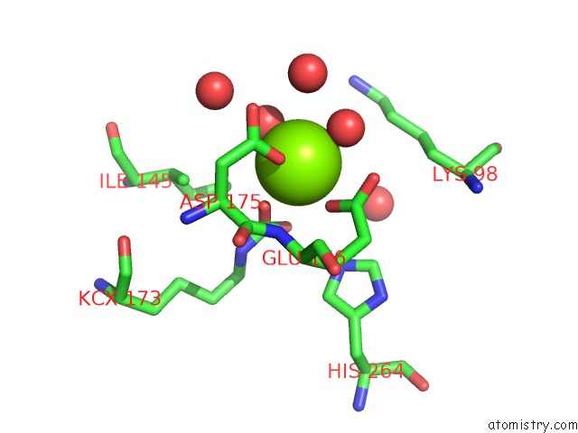
Mono view
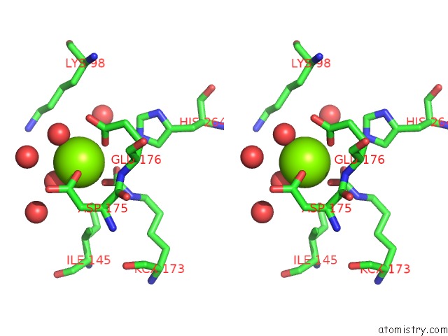
Stereo pair view
Mono view
Stereo pair view
A full contact list of Magnesium with other atoms in the Mg binding
site number 1 of Crystal Structure of A Rubisco-Like Protein From Geobacillus Kaustophilus Liganded with MG2+ Ions within 5.0Å range:
|
Magnesium binding site 2 out of 2 in 2oek
Go back to
Magnesium binding site 2 out
of 2 in the Crystal Structure of A Rubisco-Like Protein From Geobacillus Kaustophilus Liganded with MG2+ Ions
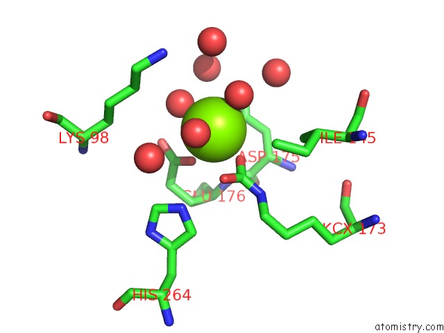
Mono view
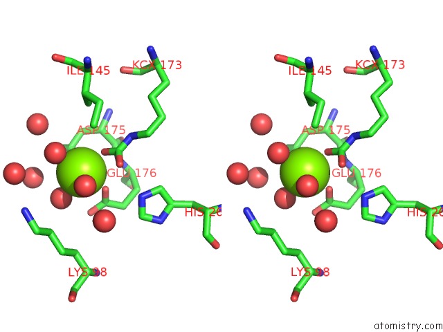
Stereo pair view
Mono view
Stereo pair view
A full contact list of Magnesium with other atoms in the Mg binding
site number 2 of Crystal Structure of A Rubisco-Like Protein From Geobacillus Kaustophilus Liganded with MG2+ Ions within 5.0Å range:
|
Reference:
H.J.Imker,
A.A.Fedorov,
E.V.Fedorov,
S.C.Almo,
J.A.Gerlt.
Mechanistic Diversity in the Rubisco Superfamily: the "Enolase" in the Methionine Salvage Pathway in Geobacillus Kaustophilus. Biochemistry V. 46 4077 2007.
ISSN: ISSN 0006-2960
PubMed: 17352497
DOI: 10.1021/BI7000483
Page generated: Wed Aug 14 01:26:22 2024
ISSN: ISSN 0006-2960
PubMed: 17352497
DOI: 10.1021/BI7000483
Last articles
Cl in 6HPXCl in 6HPF
Cl in 6HNU
Cl in 6HNR
Cl in 6HON
Cl in 6HN0
Cl in 6HN1
Cl in 6HNB
Cl in 6HNC
Cl in 6HN2