Magnesium »
PDB 2oh7-2oup »
2oi2 »
Magnesium in PDB 2oi2: Streptococcus Pneumoniae Mevalonate Kinase in Complex with Diphosphomevalonate
Enzymatic activity of Streptococcus Pneumoniae Mevalonate Kinase in Complex with Diphosphomevalonate
All present enzymatic activity of Streptococcus Pneumoniae Mevalonate Kinase in Complex with Diphosphomevalonate:
2.7.1.36;
2.7.1.36;
Protein crystallography data
The structure of Streptococcus Pneumoniae Mevalonate Kinase in Complex with Diphosphomevalonate, PDB code: 2oi2
was solved by
J.L.Andreassi,
P.W.Bilder,
M.W.Vetting,
S.L.Roderick,
T.S.Leyh,
with X-Ray Crystallography technique. A brief refinement statistics is given in the table below:
| Resolution Low / High (Å) | 30.89 / 2.50 |
| Space group | P 31 2 1 |
| Cell size a, b, c (Å), α, β, γ (°) | 101.597, 101.597, 83.280, 90.00, 90.00, 120.00 |
| R / Rfree (%) | 21.1 / 27 |
Magnesium Binding Sites:
The binding sites of Magnesium atom in the Streptococcus Pneumoniae Mevalonate Kinase in Complex with Diphosphomevalonate
(pdb code 2oi2). This binding sites where shown within
5.0 Angstroms radius around Magnesium atom.
In total only one binding site of Magnesium was determined in the Streptococcus Pneumoniae Mevalonate Kinase in Complex with Diphosphomevalonate, PDB code: 2oi2:
In total only one binding site of Magnesium was determined in the Streptococcus Pneumoniae Mevalonate Kinase in Complex with Diphosphomevalonate, PDB code: 2oi2:
Magnesium binding site 1 out of 1 in 2oi2
Go back to
Magnesium binding site 1 out
of 1 in the Streptococcus Pneumoniae Mevalonate Kinase in Complex with Diphosphomevalonate
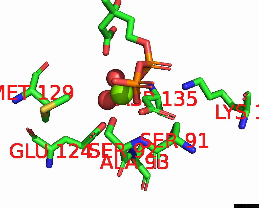
Mono view
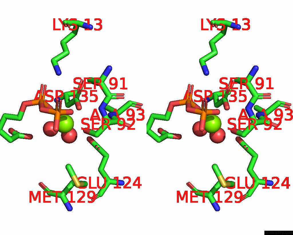
Stereo pair view
Mono view
Stereo pair view
|
|
A full contact list of Magnesium with other atoms in the Mg binding
site number 1 of Streptococcus Pneumoniae Mevalonate Kinase in Complex with Diphosphomevalonate within 5.0Å range:
|
Reference:
J.L.Andreassi,
P.W.Bilder,
M.W.Vetting,
S.L.Roderick,
T.S.Leyh.
Crystal Structure of the Streptococcus Pneumoniae Mevalonate Kinase in Complex with Diphosphomevalonate. Protein Sci. V. 16 983 2007.
ISSN: ISSN 0961-8368
PubMed: 17400916
DOI: 10.1110/PS.072755707
Page generated: Wed Aug 14 01:29:01 2024
ISSN: ISSN 0961-8368
PubMed: 17400916
DOI: 10.1110/PS.072755707
Last articles
Ca in 3V6HCa in 3V64
Ca in 3V65
Ca in 3V5U
Ca in 3V1W
Ca in 3V4F
Ca in 3V20
Ca in 3V2T
Ca in 3V13
Ca in 3V0B