Magnesium »
PDB 2oh7-2oup »
2oj6 »
Magnesium in PDB 2oj6: Crystal Structure of Reovirus T3D Attachment Protein SIGMA1 Head Domain D345N Mutant
Protein crystallography data
The structure of Crystal Structure of Reovirus T3D Attachment Protein SIGMA1 Head Domain D345N Mutant, PDB code: 2oj6
was solved by
T.Stehle,
E.Kirchner,
T.S.Dermody,
with X-Ray Crystallography technique. A brief refinement statistics is given in the table below:
| Resolution Low / High (Å) | 30.00 / 1.85 |
| Space group | P 1 21 1 |
| Cell size a, b, c (Å), α, β, γ (°) | 84.020, 51.600, 108.860, 90.00, 95.59, 90.00 |
| R / Rfree (%) | 17.5 / 22.4 |
Magnesium Binding Sites:
The binding sites of Magnesium atom in the Crystal Structure of Reovirus T3D Attachment Protein SIGMA1 Head Domain D345N Mutant
(pdb code 2oj6). This binding sites where shown within
5.0 Angstroms radius around Magnesium atom.
In total 2 binding sites of Magnesium where determined in the Crystal Structure of Reovirus T3D Attachment Protein SIGMA1 Head Domain D345N Mutant, PDB code: 2oj6:
Jump to Magnesium binding site number: 1; 2;
In total 2 binding sites of Magnesium where determined in the Crystal Structure of Reovirus T3D Attachment Protein SIGMA1 Head Domain D345N Mutant, PDB code: 2oj6:
Jump to Magnesium binding site number: 1; 2;
Magnesium binding site 1 out of 2 in 2oj6
Go back to
Magnesium binding site 1 out
of 2 in the Crystal Structure of Reovirus T3D Attachment Protein SIGMA1 Head Domain D345N Mutant
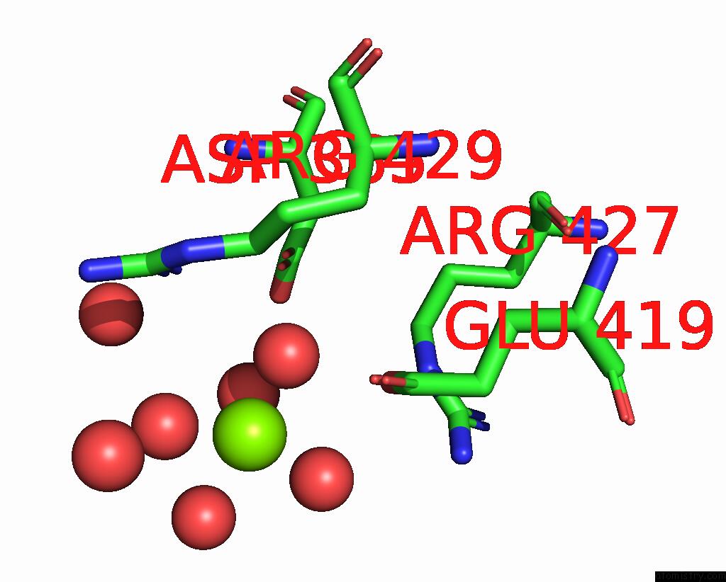
Mono view
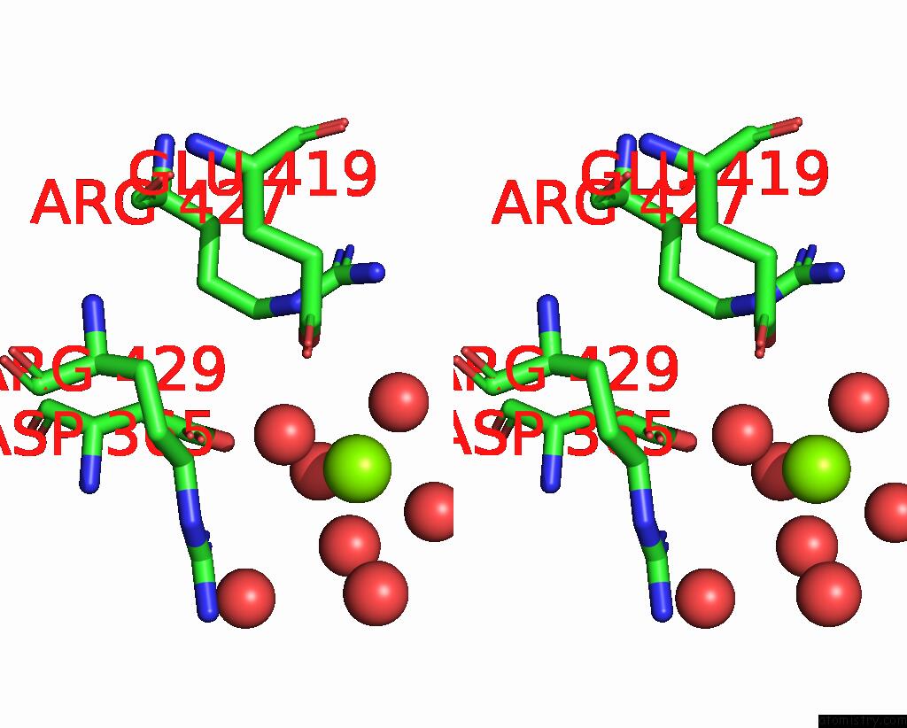
Stereo pair view
Mono view
Stereo pair view
A full contact list of Magnesium with other atoms in the Mg binding
site number 1 of Crystal Structure of Reovirus T3D Attachment Protein SIGMA1 Head Domain D345N Mutant within 5.0Å range:
|
Magnesium binding site 2 out of 2 in 2oj6
Go back to
Magnesium binding site 2 out
of 2 in the Crystal Structure of Reovirus T3D Attachment Protein SIGMA1 Head Domain D345N Mutant
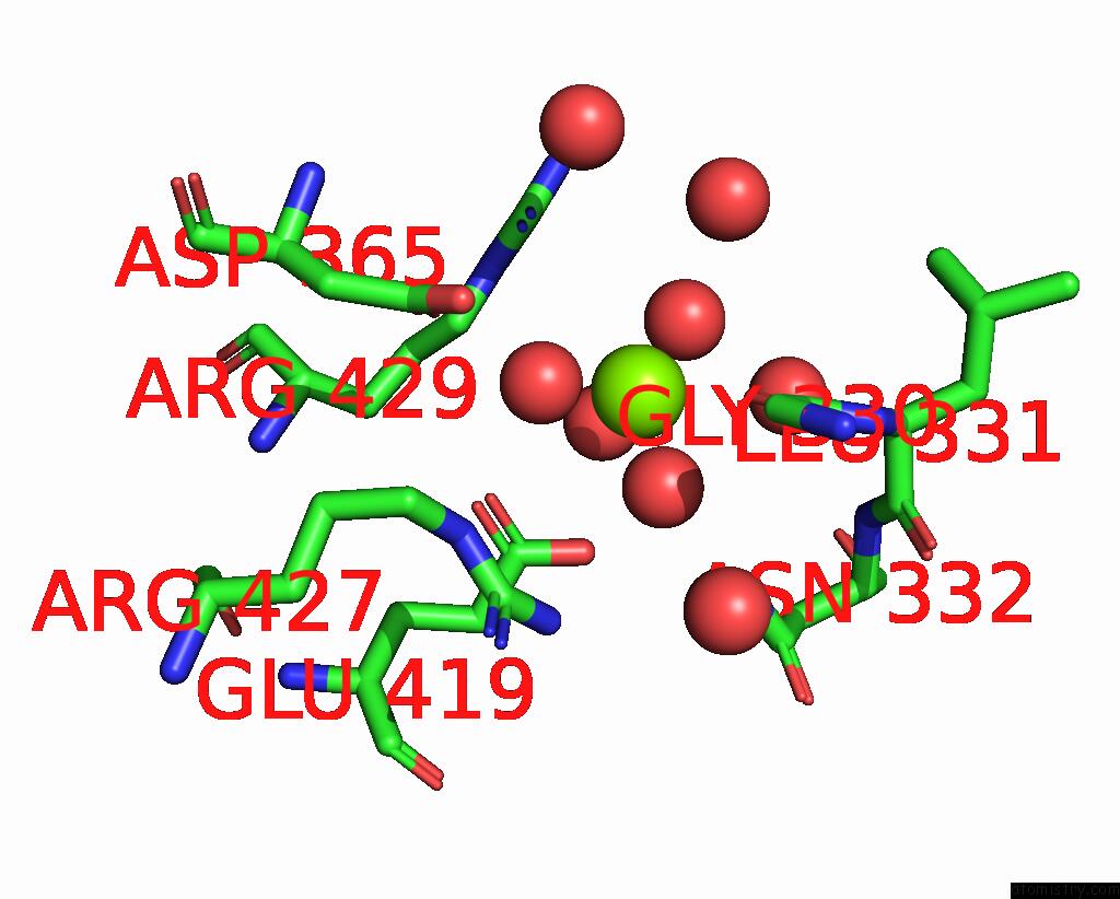
Mono view
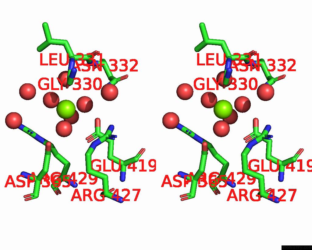
Stereo pair view
Mono view
Stereo pair view
A full contact list of Magnesium with other atoms in the Mg binding
site number 2 of Crystal Structure of Reovirus T3D Attachment Protein SIGMA1 Head Domain D345N Mutant within 5.0Å range:
|
Reference:
P.Schelling,
K.M.Guglielmi,
E.Kirchner,
B.Paetzold,
T.S.Dermody,
T.Stehle.
The Reovirus SIGMA1 Aspartic Acid Sandwich: A Trimerization Motif Poised For Conformational Change. J.Biol.Chem. V. 282 11582 2007.
ISSN: ISSN 0021-9258
PubMed: 17303562
DOI: 10.1074/JBC.M610805200
Page generated: Wed Aug 14 01:30:17 2024
ISSN: ISSN 0021-9258
PubMed: 17303562
DOI: 10.1074/JBC.M610805200
Last articles
Fe in 2YXOFe in 2YRS
Fe in 2YXC
Fe in 2YNM
Fe in 2YVJ
Fe in 2YP1
Fe in 2YU2
Fe in 2YU1
Fe in 2YQB
Fe in 2YOO