Magnesium »
PDB 2q9y-2qrf »
2qjn »
Magnesium in PDB 2qjn: Crystal Structure of D-Mannonate Dehydratase From Novosphingobium Aromaticivorans Complexed with Mg and 2-Keto-3-Deoxy-D-Gluconate
Protein crystallography data
The structure of Crystal Structure of D-Mannonate Dehydratase From Novosphingobium Aromaticivorans Complexed with Mg and 2-Keto-3-Deoxy-D-Gluconate, PDB code: 2qjn
was solved by
A.A.Fedorov,
E.V.Fedorov,
J.F.Rakus,
J.E.Vick,
J.A.Gerlt,
S.C.Almo,
with X-Ray Crystallography technique. A brief refinement statistics is given in the table below:
| Resolution Low / High (Å) | 24.89 / 2.00 |
| Space group | C 2 2 21 |
| Cell size a, b, c (Å), α, β, γ (°) | 116.269, 165.241, 167.039, 90.00, 90.00, 90.00 |
| R / Rfree (%) | 16.2 / 19.1 |
Magnesium Binding Sites:
The binding sites of Magnesium atom in the Crystal Structure of D-Mannonate Dehydratase From Novosphingobium Aromaticivorans Complexed with Mg and 2-Keto-3-Deoxy-D-Gluconate
(pdb code 2qjn). This binding sites where shown within
5.0 Angstroms radius around Magnesium atom.
In total 4 binding sites of Magnesium where determined in the Crystal Structure of D-Mannonate Dehydratase From Novosphingobium Aromaticivorans Complexed with Mg and 2-Keto-3-Deoxy-D-Gluconate, PDB code: 2qjn:
Jump to Magnesium binding site number: 1; 2; 3; 4;
In total 4 binding sites of Magnesium where determined in the Crystal Structure of D-Mannonate Dehydratase From Novosphingobium Aromaticivorans Complexed with Mg and 2-Keto-3-Deoxy-D-Gluconate, PDB code: 2qjn:
Jump to Magnesium binding site number: 1; 2; 3; 4;
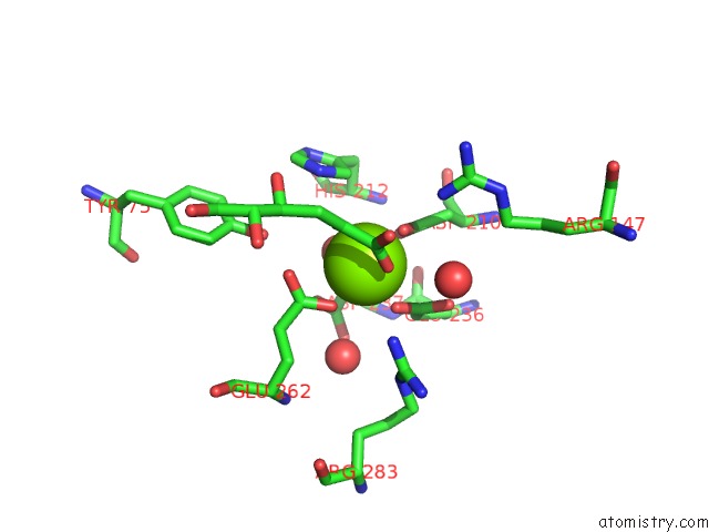
Magnesium binding site 1 out of 4 in 2qjn
Go back to
Magnesium binding site 1 out
of 4 in the Crystal Structure of D-Mannonate Dehydratase From Novosphingobium Aromaticivorans Complexed with Mg and 2-Keto-3-Deoxy-D-Gluconate
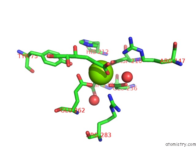
Mono view
Stereo pair view
Mono view
Stereo pair view
A full contact list of Magnesium with other atoms in the Mg binding
site number 1 of Crystal Structure of D-Mannonate Dehydratase From Novosphingobium Aromaticivorans Complexed with Mg and 2-Keto-3-Deoxy-D-Gluconate within 5.0Å range:
|
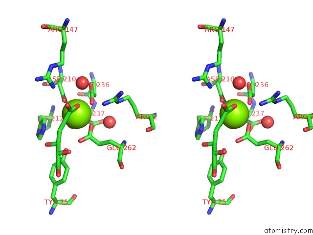
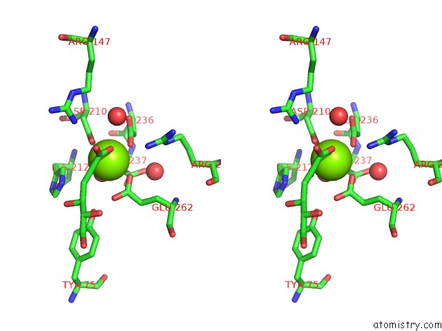
Magnesium binding site 2 out of 4 in 2qjn
Go back to
Magnesium binding site 2 out
of 4 in the Crystal Structure of D-Mannonate Dehydratase From Novosphingobium Aromaticivorans Complexed with Mg and 2-Keto-3-Deoxy-D-Gluconate
Mono view
Stereo pair view
Mono view
Stereo pair view
A full contact list of Magnesium with other atoms in the Mg binding
site number 2 of Crystal Structure of D-Mannonate Dehydratase From Novosphingobium Aromaticivorans Complexed with Mg and 2-Keto-3-Deoxy-D-Gluconate within 5.0Å range:
|
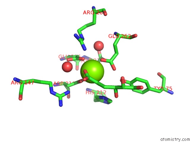
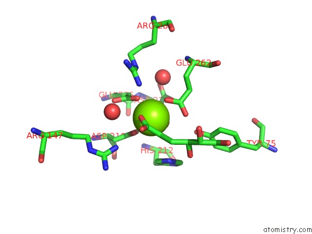
Magnesium binding site 3 out of 4 in 2qjn
Go back to
Magnesium binding site 3 out
of 4 in the Crystal Structure of D-Mannonate Dehydratase From Novosphingobium Aromaticivorans Complexed with Mg and 2-Keto-3-Deoxy-D-Gluconate
Mono view
Stereo pair view
Mono view
Stereo pair view
A full contact list of Magnesium with other atoms in the Mg binding
site number 3 of Crystal Structure of D-Mannonate Dehydratase From Novosphingobium Aromaticivorans Complexed with Mg and 2-Keto-3-Deoxy-D-Gluconate within 5.0Å range:
|
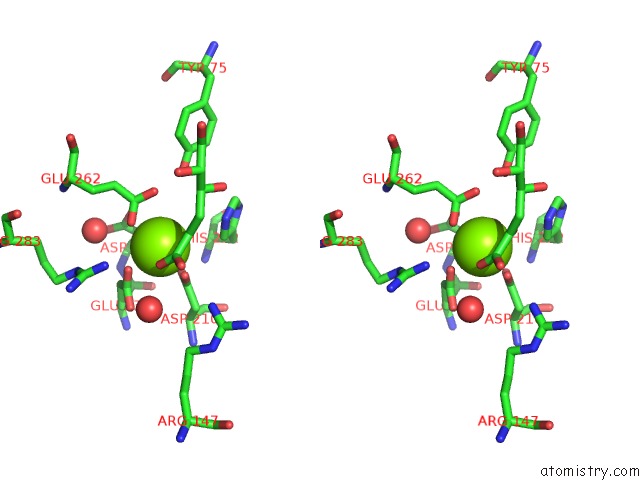
Magnesium binding site 4 out of 4 in 2qjn
Go back to
Magnesium binding site 4 out
of 4 in the Crystal Structure of D-Mannonate Dehydratase From Novosphingobium Aromaticivorans Complexed with Mg and 2-Keto-3-Deoxy-D-Gluconate
Mono view
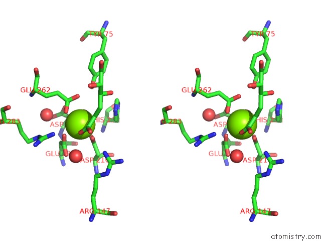
Stereo pair view
Mono view
Stereo pair view
A full contact list of Magnesium with other atoms in the Mg binding
site number 4 of Crystal Structure of D-Mannonate Dehydratase From Novosphingobium Aromaticivorans Complexed with Mg and 2-Keto-3-Deoxy-D-Gluconate within 5.0Å range:
|
Reference:
J.F.Rakus,
A.A.Fedorov,
E.V.Fedorov,
M.E.Glasner,
J.E.Vick,
P.C.Babbitt,
S.C.Almo,
J.A.Gerlt.
Evolution of Enzymatic Activities in the Enolase Superfamily: D-Mannonate Dehydratase From Novosphingobium Aromaticivorans. Biochemistry V. 46 12896 2007.
ISSN: ISSN 0006-2960
PubMed: 17944491
DOI: 10.1021/BI701703W
Page generated: Sun Aug 10 13:22:10 2025
ISSN: ISSN 0006-2960
PubMed: 17944491
DOI: 10.1021/BI701703W
Last articles
Mg in 4EPYMg in 4EPX
Mg in 4EPW
Mg in 4EPV
Mg in 4EPT
Mg in 4EPR
Mg in 4EPK
Mg in 4EOQ
Mg in 4EOY
Mg in 4EP4