Magnesium »
PDB 2qrn-2r1y »
2qs8 »
Magnesium in PDB 2qs8: Crystal Structure of A Xaa-Pro Dipeptidase with Bound Methionine in the Active Site
Protein crystallography data
The structure of Crystal Structure of A Xaa-Pro Dipeptidase with Bound Methionine in the Active Site, PDB code: 2qs8
was solved by
D.Kumaran,
S.K.Burley,
S.Swaminathan,
New York Sgx Research Center Forstructural Genomics (Nysgxrc),
with X-Ray Crystallography technique. A brief refinement statistics is given in the table below:
| Resolution Low / High (Å) | 45.81 / 2.33 |
| Space group | I 4 |
| Cell size a, b, c (Å), α, β, γ (°) | 144.846, 144.846, 101.029, 90.00, 90.00, 90.00 |
| R / Rfree (%) | 20.7 / 23.4 |
Magnesium Binding Sites:
The binding sites of Magnesium atom in the Crystal Structure of A Xaa-Pro Dipeptidase with Bound Methionine in the Active Site
(pdb code 2qs8). This binding sites where shown within
5.0 Angstroms radius around Magnesium atom.
In total 2 binding sites of Magnesium where determined in the Crystal Structure of A Xaa-Pro Dipeptidase with Bound Methionine in the Active Site, PDB code: 2qs8:
Jump to Magnesium binding site number: 1; 2;
In total 2 binding sites of Magnesium where determined in the Crystal Structure of A Xaa-Pro Dipeptidase with Bound Methionine in the Active Site, PDB code: 2qs8:
Jump to Magnesium binding site number: 1; 2;
Magnesium binding site 1 out of 2 in 2qs8
Go back to
Magnesium binding site 1 out
of 2 in the Crystal Structure of A Xaa-Pro Dipeptidase with Bound Methionine in the Active Site
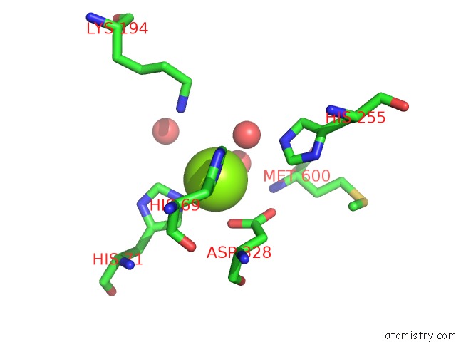
Mono view
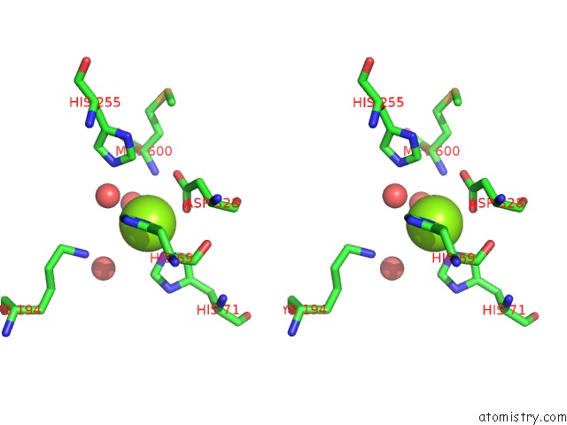
Stereo pair view
Mono view
Stereo pair view
A full contact list of Magnesium with other atoms in the Mg binding
site number 1 of Crystal Structure of A Xaa-Pro Dipeptidase with Bound Methionine in the Active Site within 5.0Å range:
|
Magnesium binding site 2 out of 2 in 2qs8
Go back to
Magnesium binding site 2 out
of 2 in the Crystal Structure of A Xaa-Pro Dipeptidase with Bound Methionine in the Active Site
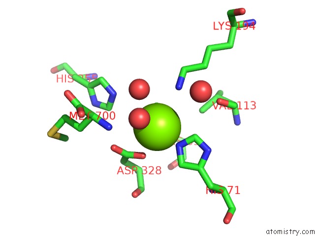
Mono view
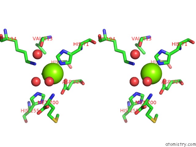
Stereo pair view
Mono view
Stereo pair view
A full contact list of Magnesium with other atoms in the Mg binding
site number 2 of Crystal Structure of A Xaa-Pro Dipeptidase with Bound Methionine in the Active Site within 5.0Å range:
|
Reference:
D.F.Xiang,
C.Xu,
D.Kumaran,
A.C.Brown,
J.M.Sauder,
S.K.Burley,
S.Swaminathan,
F.M.Raushel.
Functional Annotation of Two New Carboxypeptidases From the Amidohydrolase Superfamily of Enzymes. Biochemistry V. 48 4567 2009.
ISSN: ISSN 0006-2960
PubMed: 19358546
DOI: 10.1021/BI900453U
Page generated: Sun Aug 10 13:36:07 2025
ISSN: ISSN 0006-2960
PubMed: 19358546
DOI: 10.1021/BI900453U
Last articles
Mn in 9LJUMn in 9LJW
Mn in 9LJS
Mn in 9LJR
Mn in 9LJT
Mn in 9LJV
Mg in 9UA2
Mg in 9R96
Mg in 9VM1
Mg in 9P01