Magnesium »
PDB 2r20-2reu »
2r20 »
Magnesium in PDB 2r20: Structure of the Rna Brominated Tridecamer R(Gcguu- 5BUGAAACGC) at 1.3 A (BR2)
Protein crystallography data
The structure of Structure of the Rna Brominated Tridecamer R(Gcguu- 5BUGAAACGC) at 1.3 A (BR2), PDB code: 2r20
was solved by
Y.Timsit,
S.Bombard,
with X-Ray Crystallography technique. A brief refinement statistics is given in the table below:
| Resolution Low / High (Å) | 20.00 / 1.30 |
| Space group | P 1 |
| Cell size a, b, c (Å), α, β, γ (°) | 25.280, 25.800, 29.990, 98.62, 105.58, 100.92 |
| R / Rfree (%) | 18.1 / 19.5 |
Other elements in 2r20:
The structure of Structure of the Rna Brominated Tridecamer R(Gcguu- 5BUGAAACGC) at 1.3 A (BR2) also contains other interesting chemical elements:
| Bromine | (Br) | 2 atoms |
| Sodium | (Na) | 5 atoms |
Magnesium Binding Sites:
The binding sites of Magnesium atom in the Structure of the Rna Brominated Tridecamer R(Gcguu- 5BUGAAACGC) at 1.3 A (BR2)
(pdb code 2r20). This binding sites where shown within
5.0 Angstroms radius around Magnesium atom.
In total only one binding site of Magnesium was determined in the Structure of the Rna Brominated Tridecamer R(Gcguu- 5BUGAAACGC) at 1.3 A (BR2), PDB code: 2r20:
In total only one binding site of Magnesium was determined in the Structure of the Rna Brominated Tridecamer R(Gcguu- 5BUGAAACGC) at 1.3 A (BR2), PDB code: 2r20:
Magnesium binding site 1 out of 1 in 2r20
Go back to
Magnesium binding site 1 out
of 1 in the Structure of the Rna Brominated Tridecamer R(Gcguu- 5BUGAAACGC) at 1.3 A (BR2)
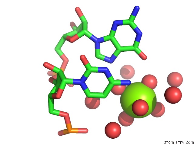
Mono view
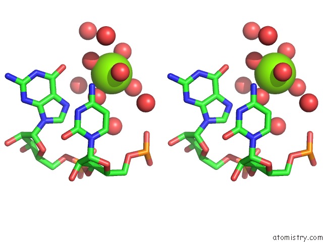
Stereo pair view
Mono view
Stereo pair view
A full contact list of Magnesium with other atoms in the Mg binding
site number 1 of Structure of the Rna Brominated Tridecamer R(Gcguu- 5BUGAAACGC) at 1.3 A (BR2) within 5.0Å range:
|
Reference:
Y.Timsit,
S.Bombard.
The 1.3 A Resolution Structure of the Rna Tridecamer R(Gcguuugaaacgc): Metal Ion Binding Correlates with Base Unstacking and Groove Contraction. Rna V. 13 2098 2007.
ISSN: ISSN 1355-8382
PubMed: 17940138
DOI: 10.1261/RNA.730207
Page generated: Sun Aug 10 13:41:19 2025
ISSN: ISSN 1355-8382
PubMed: 17940138
DOI: 10.1261/RNA.730207
Last articles
Mg in 2UU7Mg in 2UAG
Mg in 2UKD
Mg in 2SHK
Mg in 2TPS
Mg in 2TRT
Mg in 2TRA
Mg in 2RMK
Mg in 2RUS
Mg in 2TCT