Magnesium »
PDB 2rex-2uxd »
2rfg »
Magnesium in PDB 2rfg: Crystal Structure of Dihydrodipicolinate Synthase From Hahella Chejuensis at 1.5A Resolution
Enzymatic activity of Crystal Structure of Dihydrodipicolinate Synthase From Hahella Chejuensis at 1.5A Resolution
All present enzymatic activity of Crystal Structure of Dihydrodipicolinate Synthase From Hahella Chejuensis at 1.5A Resolution:
4.2.1.52;
4.2.1.52;
Protein crystallography data
The structure of Crystal Structure of Dihydrodipicolinate Synthase From Hahella Chejuensis at 1.5A Resolution, PDB code: 2rfg
was solved by
B.S.Kang,
M.H.Kim,
G.H.Kim,
K.J.Kim,
with X-Ray Crystallography technique. A brief refinement statistics is given in the table below:
| Resolution Low / High (Å) | 19.98 / 1.50 |
| Space group | P 21 21 21 |
| Cell size a, b, c (Å), α, β, γ (°) | 67.061, 120.644, 161.137, 90.00, 90.00, 90.00 |
| R / Rfree (%) | 15.9 / 17.8 |
Magnesium Binding Sites:
The binding sites of Magnesium atom in the Crystal Structure of Dihydrodipicolinate Synthase From Hahella Chejuensis at 1.5A Resolution
(pdb code 2rfg). This binding sites where shown within
5.0 Angstroms radius around Magnesium atom.
In total only one binding site of Magnesium was determined in the Crystal Structure of Dihydrodipicolinate Synthase From Hahella Chejuensis at 1.5A Resolution, PDB code: 2rfg:
In total only one binding site of Magnesium was determined in the Crystal Structure of Dihydrodipicolinate Synthase From Hahella Chejuensis at 1.5A Resolution, PDB code: 2rfg:
Magnesium binding site 1 out of 1 in 2rfg
Go back to
Magnesium binding site 1 out
of 1 in the Crystal Structure of Dihydrodipicolinate Synthase From Hahella Chejuensis at 1.5A Resolution
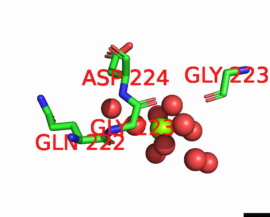
Mono view
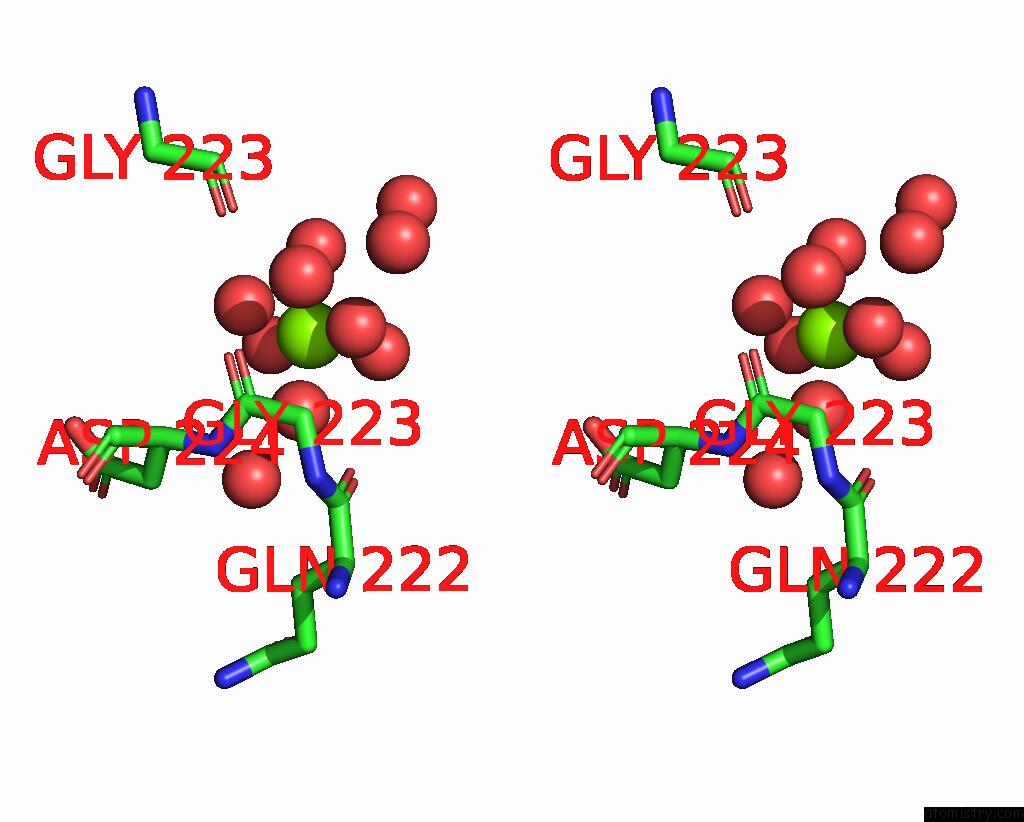
Stereo pair view
Mono view
Stereo pair view
A full contact list of Magnesium with other atoms in the Mg binding
site number 1 of Crystal Structure of Dihydrodipicolinate Synthase From Hahella Chejuensis at 1.5A Resolution within 5.0Å range:
|
Reference:
B.S.Kang,
M.H.Kim,
G.H.Kim,
K.J.Kim.
Crystal Structure of Dihydrodipicolinate Synthase From Hahella Chejuensis at 1.5A Resolution To Be Published.
Page generated: Sun Aug 10 13:46:20 2025
Last articles
Mg in 2WTZMg in 2WTY
Mg in 2WTP
Mg in 2WTO
Mg in 2WSS
Mg in 2WSB
Mg in 2WPD
Mg in 2WOQ
Mg in 2WOJ
Mg in 2WQS