Magnesium »
PDB 2uxj-2v7y »
2uyr »
Magnesium in PDB 2uyr: Crystal Structure of Bacillus Cereus Sphingomyelinase Mutant :N57A
Enzymatic activity of Crystal Structure of Bacillus Cereus Sphingomyelinase Mutant :N57A
All present enzymatic activity of Crystal Structure of Bacillus Cereus Sphingomyelinase Mutant :N57A:
3.1.4.12;
3.1.4.12;
Protein crystallography data
The structure of Crystal Structure of Bacillus Cereus Sphingomyelinase Mutant :N57A, PDB code: 2uyr
was solved by
M.Oda,
H.Tsuge,
J.Sakurai,
with X-Ray Crystallography technique. A brief refinement statistics is given in the table below:
| Resolution Low / High (Å) | 54.15 / 2.40 |
| Space group | P 21 21 21 |
| Cell size a, b, c (Å), α, β, γ (°) | 55.090, 63.927, 101.690, 90.00, 90.00, 90.00 |
| R / Rfree (%) | 18.2 / 23.5 |
Magnesium Binding Sites:
The binding sites of Magnesium atom in the Crystal Structure of Bacillus Cereus Sphingomyelinase Mutant :N57A
(pdb code 2uyr). This binding sites where shown within
5.0 Angstroms radius around Magnesium atom.
In total only one binding site of Magnesium was determined in the Crystal Structure of Bacillus Cereus Sphingomyelinase Mutant :N57A, PDB code: 2uyr:
In total only one binding site of Magnesium was determined in the Crystal Structure of Bacillus Cereus Sphingomyelinase Mutant :N57A, PDB code: 2uyr:
Magnesium binding site 1 out of 1 in 2uyr
Go back to
Magnesium binding site 1 out
of 1 in the Crystal Structure of Bacillus Cereus Sphingomyelinase Mutant :N57A
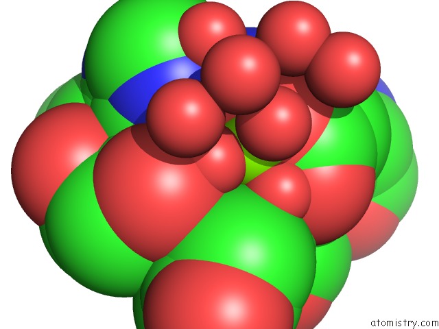
Mono view
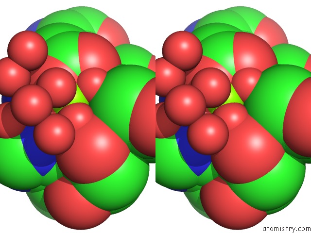
Stereo pair view
Mono view
Stereo pair view
A full contact list of Magnesium with other atoms in the Mg binding
site number 1 of Crystal Structure of Bacillus Cereus Sphingomyelinase Mutant :N57A within 5.0Å range:
|
Reference:
M.Oda,
H.Tsuge,
J.Sakurai.
Crystal Structure of Bacillus Cereus Sphingomyelinase Mutant : N57A To Be Published.
Page generated: Wed Aug 14 04:56:08 2024
Last articles
Mg in 1IKKMg in 1IK5
Mg in 1IJJ
Mg in 1IJF
Mg in 1IJD
Mg in 1IIR
Mg in 1II9
Mg in 1II6
Mg in 1II0
Mg in 1IHU