Magnesium »
PDB 2uxj-2v7y »
2v0y »
Magnesium in PDB 2v0y: Crystal Structure of Apo C298S Tryptophanase From E.Coli
Enzymatic activity of Crystal Structure of Apo C298S Tryptophanase From E.Coli
All present enzymatic activity of Crystal Structure of Apo C298S Tryptophanase From E.Coli:
4.1.99.1;
4.1.99.1;
Protein crystallography data
The structure of Crystal Structure of Apo C298S Tryptophanase From E.Coli, PDB code: 2v0y
was solved by
A.Kogan,
G.Y.Gdalevsky,
R.Cohen-Luria,
Y.Goldgur,
A.H.Parola,
O.Almog,
with X-Ray Crystallography technique. A brief refinement statistics is given in the table below:
| Resolution Low / High (Å) | 80.00 / 2.00 |
| Space group | F 2 2 2 |
| Cell size a, b, c (Å), α, β, γ (°) | 120.484, 118.770, 171.534, 90.00, 90.00, 90.00 |
| R / Rfree (%) | 21.5 / 25.7 |
Other elements in 2v0y:
The structure of Crystal Structure of Apo C298S Tryptophanase From E.Coli also contains other interesting chemical elements:
| Chlorine | (Cl) | 1 atom |
Magnesium Binding Sites:
The binding sites of Magnesium atom in the Crystal Structure of Apo C298S Tryptophanase From E.Coli
(pdb code 2v0y). This binding sites where shown within
5.0 Angstroms radius around Magnesium atom.
In total only one binding site of Magnesium was determined in the Crystal Structure of Apo C298S Tryptophanase From E.Coli, PDB code: 2v0y:
In total only one binding site of Magnesium was determined in the Crystal Structure of Apo C298S Tryptophanase From E.Coli, PDB code: 2v0y:
Magnesium binding site 1 out of 1 in 2v0y
Go back to
Magnesium binding site 1 out
of 1 in the Crystal Structure of Apo C298S Tryptophanase From E.Coli
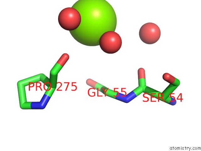
Mono view
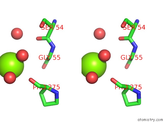
Stereo pair view
Mono view
Stereo pair view
A full contact list of Magnesium with other atoms in the Mg binding
site number 1 of Crystal Structure of Apo C298S Tryptophanase From E.Coli within 5.0Å range:
|
Reference:
A.Kogan,
G.Y.Gdalevsky,
R.Cohen-Luria,
Y.Goldgur,
R.S.Phillips,
A.H.Parola,
O.Almog.
Conformational Changes and Loose Packing Promote E. Coli Tryptophanase Cold Lability. Bmc Struct.Biol. V. 9 65 2009.
ISSN: ESSN 1472-6807
PubMed: 19814824
DOI: 10.1186/1472-6807-9-65
Page generated: Wed Aug 14 04:56:57 2024
ISSN: ESSN 1472-6807
PubMed: 19814824
DOI: 10.1186/1472-6807-9-65
Last articles
Mg in 1RTDMg in 1RTK
Mg in 1RRP
Mg in 1RS0
Mg in 1RRG
Mg in 1RRF
Mg in 1RQJ
Mg in 1RQY
Mg in 1RQN
Mg in 1RQI