Magnesium »
PDB 2v9j-2vfj »
2ve9 »
Magnesium in PDB 2ve9: Xray Structure of Kops Bound Gamma Domain of Ftsk (P. Aeruginosa)
Protein crystallography data
The structure of Xray Structure of Kops Bound Gamma Domain of Ftsk (P. Aeruginosa), PDB code: 2ve9
was solved by
J.Lowe,
M.D.Allen,
D.J.Sherratt,
with X-Ray Crystallography technique. A brief refinement statistics is given in the table below:
| Resolution Low / High (Å) | 66.67 / 1.9 |
| Space group | C 1 2 1 |
| Cell size a, b, c (Å), α, β, γ (°) | 137.937, 63.073, 76.026, 90.00, 118.76, 90.00 |
| R / Rfree (%) | 19.6 / 24.9 |
Magnesium Binding Sites:
The binding sites of Magnesium atom in the Xray Structure of Kops Bound Gamma Domain of Ftsk (P. Aeruginosa)
(pdb code 2ve9). This binding sites where shown within
5.0 Angstroms radius around Magnesium atom.
In total only one binding site of Magnesium was determined in the Xray Structure of Kops Bound Gamma Domain of Ftsk (P. Aeruginosa), PDB code: 2ve9:
In total only one binding site of Magnesium was determined in the Xray Structure of Kops Bound Gamma Domain of Ftsk (P. Aeruginosa), PDB code: 2ve9:
Magnesium binding site 1 out of 1 in 2ve9
Go back to
Magnesium binding site 1 out
of 1 in the Xray Structure of Kops Bound Gamma Domain of Ftsk (P. Aeruginosa)
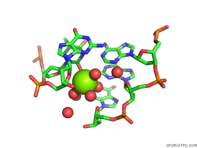
Mono view
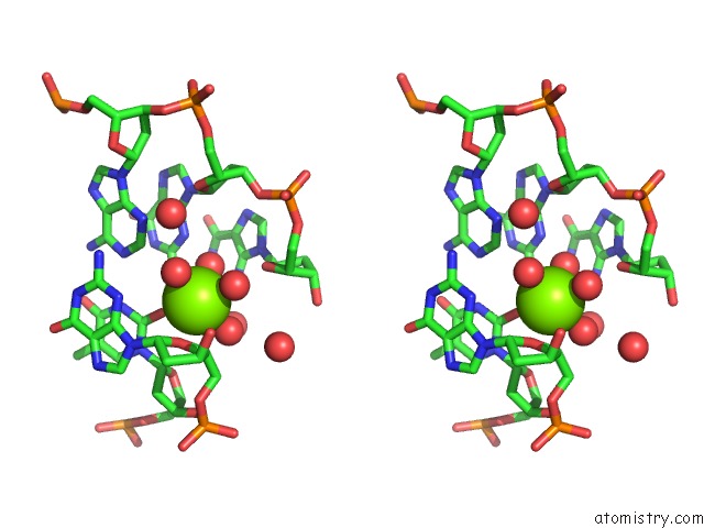
Stereo pair view
Mono view
Stereo pair view
A full contact list of Magnesium with other atoms in the Mg binding
site number 1 of Xray Structure of Kops Bound Gamma Domain of Ftsk (P. Aeruginosa) within 5.0Å range:
|
Reference:
J.Lowe,
A.Ellonen,
M.D.Allen,
C.Atkinson,
D.J.Sherratt,
I.Grainge.
Molecular Mechanism of Sequence-Directed Dna Loading and Translocation By Ftsk. Mol.Cell V. 31 498 2008.
ISSN: ISSN 1097-2765
PubMed: 18722176
DOI: 10.1016/J.MOLCEL.2008.05.027
Page generated: Wed Aug 14 05:10:36 2024
ISSN: ISSN 1097-2765
PubMed: 18722176
DOI: 10.1016/J.MOLCEL.2008.05.027
Last articles
Mg in 2UAGMg in 2UKD
Mg in 2SHK
Mg in 2TPS
Mg in 2TRT
Mg in 2TRA
Mg in 2RMK
Mg in 2RUS
Mg in 2TCT
Mg in 2RHD