Magnesium »
PDB 2x13-2xbn »
2x20 »
Magnesium in PDB 2x20: Structure of Peridinin-Chlorophyll-Protein Reconstituted with Chl-B
Protein crystallography data
The structure of Structure of Peridinin-Chlorophyll-Protein Reconstituted with Chl-B, PDB code: 2x20
was solved by
T.Schulte,
R.G.Hiller,
E.Hofmann,
with X-Ray Crystallography technique. A brief refinement statistics is given in the table below:
| Resolution Low / High (Å) | 43.15 / 1.95 |
| Space group | C 2 2 21 |
| Cell size a, b, c (Å), α, β, γ (°) | 68.570, 82.000, 75.410, 90.00, 90.00, 90.00 |
| R / Rfree (%) | 15.5 / 19.6 |
Other elements in 2x20:
The structure of Structure of Peridinin-Chlorophyll-Protein Reconstituted with Chl-B also contains other interesting chemical elements:
| Potassium | (K) | 1 atom |
| Cadmium | (Cd) | 8 atoms |
| Chlorine | (Cl) | 3 atoms |
| Sodium | (Na) | 1 atom |
Magnesium Binding Sites:
The binding sites of Magnesium atom in the Structure of Peridinin-Chlorophyll-Protein Reconstituted with Chl-B
(pdb code 2x20). This binding sites where shown within
5.0 Angstroms radius around Magnesium atom.
In total only one binding site of Magnesium was determined in the Structure of Peridinin-Chlorophyll-Protein Reconstituted with Chl-B, PDB code: 2x20:
In total only one binding site of Magnesium was determined in the Structure of Peridinin-Chlorophyll-Protein Reconstituted with Chl-B, PDB code: 2x20:
Magnesium binding site 1 out of 1 in 2x20
Go back to
Magnesium binding site 1 out
of 1 in the Structure of Peridinin-Chlorophyll-Protein Reconstituted with Chl-B
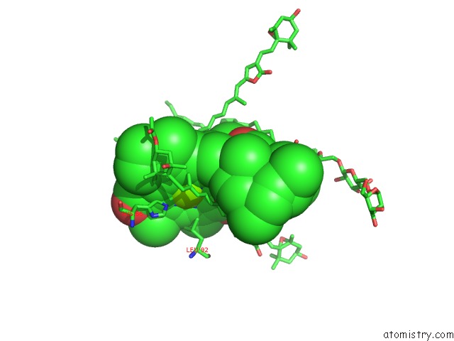
Mono view
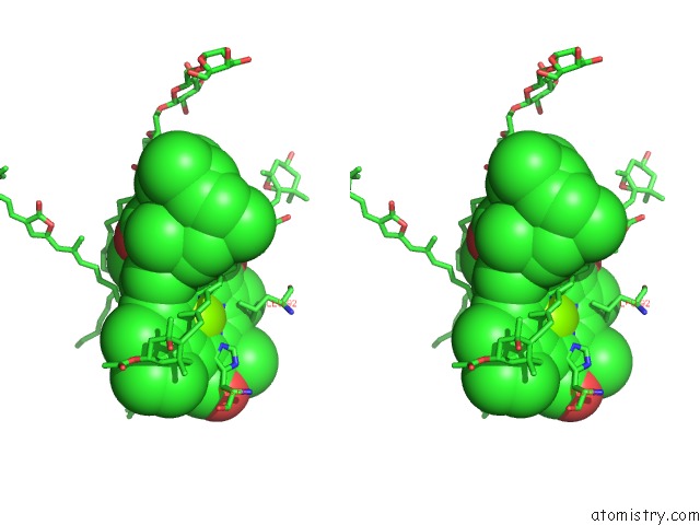
Stereo pair view
Mono view
Stereo pair view
A full contact list of Magnesium with other atoms in the Mg binding
site number 1 of Structure of Peridinin-Chlorophyll-Protein Reconstituted with Chl-B within 5.0Å range:
|
Reference:
T.Schulte,
R.G.Hiller,
E.Hofmann.
X-Ray Structures of the Peridinin-Chlorophyll-Protein Reconstituted with Different Chlorophylls. Febs Lett. V. 584 973 2010.
ISSN: ISSN 0014-5793
PubMed: 20102711
DOI: 10.1016/J.FEBSLET.2010.01.041
Page generated: Sun Aug 10 16:18:17 2025
ISSN: ISSN 0014-5793
PubMed: 20102711
DOI: 10.1016/J.FEBSLET.2010.01.041
Last articles
Mn in 5TBBMn in 5T7P
Mn in 5T4I
Mn in 5T3V
Mn in 5T30
Mn in 5SVC
Mn in 5T2T
Mn in 5SVB
Mn in 5SSV
Mn in 5SSW