Magnesium »
PDB 2xni-2xzs »
2xo8 »
Magnesium in PDB 2xo8: Crystal Structure of Myosin-2 in Complex with Tribromodichloropseudilin
Protein crystallography data
The structure of Crystal Structure of Myosin-2 in Complex with Tribromodichloropseudilin, PDB code: 2xo8
was solved by
M.Preller,
K.Chinthalapudi,
R.Martin,
H.J.Knoelker,
D.J.Manstein,
with X-Ray Crystallography technique. A brief refinement statistics is given in the table below:
| Resolution Low / High (Å) | 19.70 / 2.40 |
| Space group | C 2 2 21 |
| Cell size a, b, c (Å), α, β, γ (°) | 88.340, 146.383, 152.798, 90.00, 90.00, 90.00 |
| R / Rfree (%) | 20.2 / 25.4 |
Other elements in 2xo8:
The structure of Crystal Structure of Myosin-2 in Complex with Tribromodichloropseudilin also contains other interesting chemical elements:
| Chlorine | (Cl) | 2 atoms |
| Bromine | (Br) | 3 atoms |
| Vanadium | (V) | 1 atom |
Magnesium Binding Sites:
The binding sites of Magnesium atom in the Crystal Structure of Myosin-2 in Complex with Tribromodichloropseudilin
(pdb code 2xo8). This binding sites where shown within
5.0 Angstroms radius around Magnesium atom.
In total only one binding site of Magnesium was determined in the Crystal Structure of Myosin-2 in Complex with Tribromodichloropseudilin, PDB code: 2xo8:
In total only one binding site of Magnesium was determined in the Crystal Structure of Myosin-2 in Complex with Tribromodichloropseudilin, PDB code: 2xo8:
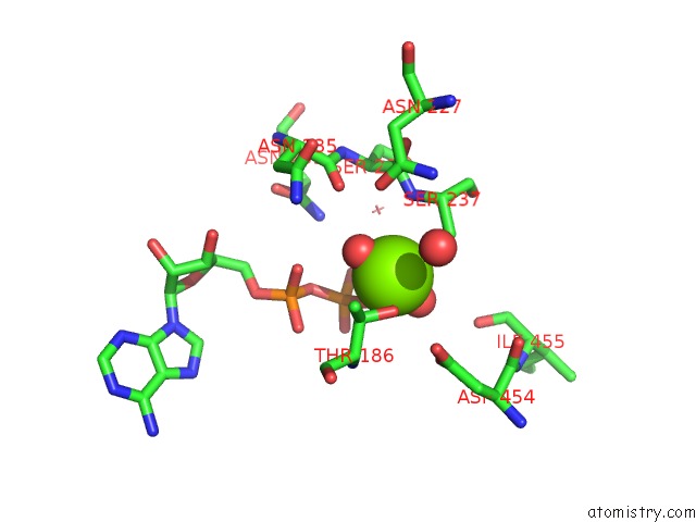
Magnesium binding site 1 out of 1 in 2xo8
Go back to
Magnesium binding site 1 out
of 1 in the Crystal Structure of Myosin-2 in Complex with Tribromodichloropseudilin
Mono view

Stereo pair view
Mono view
Stereo pair view
A full contact list of Magnesium with other atoms in the Mg binding
site number 1 of Crystal Structure of Myosin-2 in Complex with Tribromodichloropseudilin within 5.0Å range:
|
Reference:
M.Preller,
K.Chinthalapudi,
R.Martin,
H.Knolker,
D.J.Manstein.
Inhibition of Myosin Atpase Activity By Halogenated Pseudilins: A Structure-Activity Study. J.Med.Chem. V. 54 3675 2011.
ISSN: ISSN 0022-2623
PubMed: 21534527
DOI: 10.1021/JM200259F
Page generated: Sun Aug 10 16:30:37 2025
ISSN: ISSN 0022-2623
PubMed: 21534527
DOI: 10.1021/JM200259F
Last articles
Mg in 3HWXMg in 3HWO
Mg in 3HWW
Mg in 3HWT
Mg in 3HW5
Mg in 3HWS
Mg in 3HQP
Mg in 3HW8
Mg in 3HVR
Mg in 3HW4