Magnesium »
PDB 2xni-2xzs »
2xsx »
Magnesium in PDB 2xsx: Crystal Structure of Human Beta Enolase Enob
Enzymatic activity of Crystal Structure of Human Beta Enolase Enob
All present enzymatic activity of Crystal Structure of Human Beta Enolase Enob:
4.2.1.11;
4.2.1.11;
Protein crystallography data
The structure of Crystal Structure of Human Beta Enolase Enob, PDB code: 2xsx
was solved by
M.Vollmar,
E.Krysztofinska,
A.Chaikuad,
T.Krojer,
R.Cocking,
F.Von Delft,
C.Bountra,
C.H.Arrowsmith,
J.Weigelt,
A.Edwards,
W.W.Yue,
U.Oppermann,
with X-Ray Crystallography technique. A brief refinement statistics is given in the table below:
| Resolution Low / High (Å) | 29.39 / 1.70 |
| Space group | P 21 21 21 |
| Cell size a, b, c (Å), α, β, γ (°) | 95.878, 105.531, 106.091, 90.00, 90.00, 90.00 |
| R / Rfree (%) | 15.3 / 18.9 |
Magnesium Binding Sites:
The binding sites of Magnesium atom in the Crystal Structure of Human Beta Enolase Enob
(pdb code 2xsx). This binding sites where shown within
5.0 Angstroms radius around Magnesium atom.
In total 2 binding sites of Magnesium where determined in the Crystal Structure of Human Beta Enolase Enob, PDB code: 2xsx:
Jump to Magnesium binding site number: 1; 2;
In total 2 binding sites of Magnesium where determined in the Crystal Structure of Human Beta Enolase Enob, PDB code: 2xsx:
Jump to Magnesium binding site number: 1; 2;
Magnesium binding site 1 out of 2 in 2xsx
Go back to
Magnesium binding site 1 out
of 2 in the Crystal Structure of Human Beta Enolase Enob
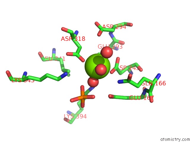
Mono view
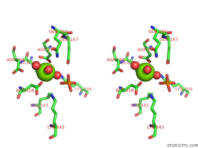
Stereo pair view
Mono view
Stereo pair view
A full contact list of Magnesium with other atoms in the Mg binding
site number 1 of Crystal Structure of Human Beta Enolase Enob within 5.0Å range:
|
Magnesium binding site 2 out of 2 in 2xsx
Go back to
Magnesium binding site 2 out
of 2 in the Crystal Structure of Human Beta Enolase Enob
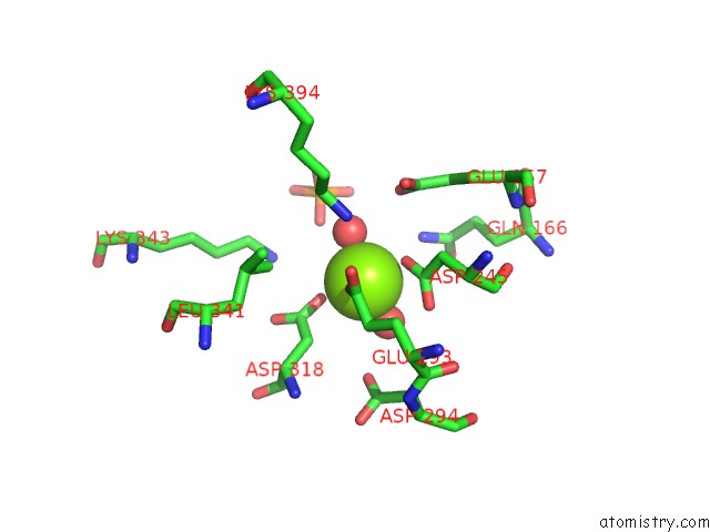
Mono view
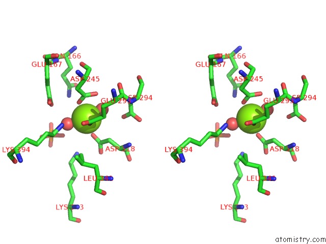
Stereo pair view
Mono view
Stereo pair view
A full contact list of Magnesium with other atoms in the Mg binding
site number 2 of Crystal Structure of Human Beta Enolase Enob within 5.0Å range:
|
Reference:
M.Vollmar,
E.Krysztofinska,
A.Chaikuad,
T.Krojer,
R.Cocking,
F.Von Delft,
C.Bountra,
C.H.Arrowsmith,
J.Weigelt,
A.Edwards,
W.W.Yue,
U.Oppermann.
Crystal Structure of Human Beta Enolase Enob To Be Published.
Page generated: Sun Aug 10 16:31:54 2025
Last articles
Mg in 4KGGMg in 4KG3
Mg in 4KGD
Mg in 4KGC
Mg in 4KG0
Mg in 4KFT
Mg in 4KFU
Mg in 4KFJ
Mg in 4KFS
Mg in 4KFR