Magnesium »
PDB 2ynj-2z4w »
2yxu »
Magnesium in PDB 2yxu: Human Pyridoxal Kinase
Enzymatic activity of Human Pyridoxal Kinase
All present enzymatic activity of Human Pyridoxal Kinase:
2.7.1.35;
2.7.1.35;
Protein crystallography data
The structure of Human Pyridoxal Kinase, PDB code: 2yxu
was solved by
M.K.Safo,
F.N.Musayev,
T.P.Ko,
V.Schirch,
with X-Ray Crystallography technique. A brief refinement statistics is given in the table below:
| Resolution Low / High (Å) | 30.00 / 2.20 |
| Space group | I 2 2 2 |
| Cell size a, b, c (Å), α, β, γ (°) | 90.462, 115.119, 170.201, 90.00, 90.00, 90.00 |
| R / Rfree (%) | 19.2 / 24.3 |
Other elements in 2yxu:
The structure of Human Pyridoxal Kinase also contains other interesting chemical elements:
| Sodium | (Na) | 2 atoms |
Magnesium Binding Sites:
The binding sites of Magnesium atom in the Human Pyridoxal Kinase
(pdb code 2yxu). This binding sites where shown within
5.0 Angstroms radius around Magnesium atom.
In total 2 binding sites of Magnesium where determined in the Human Pyridoxal Kinase, PDB code: 2yxu:
Jump to Magnesium binding site number: 1; 2;
In total 2 binding sites of Magnesium where determined in the Human Pyridoxal Kinase, PDB code: 2yxu:
Jump to Magnesium binding site number: 1; 2;
Magnesium binding site 1 out of 2 in 2yxu
Go back to
Magnesium binding site 1 out
of 2 in the Human Pyridoxal Kinase
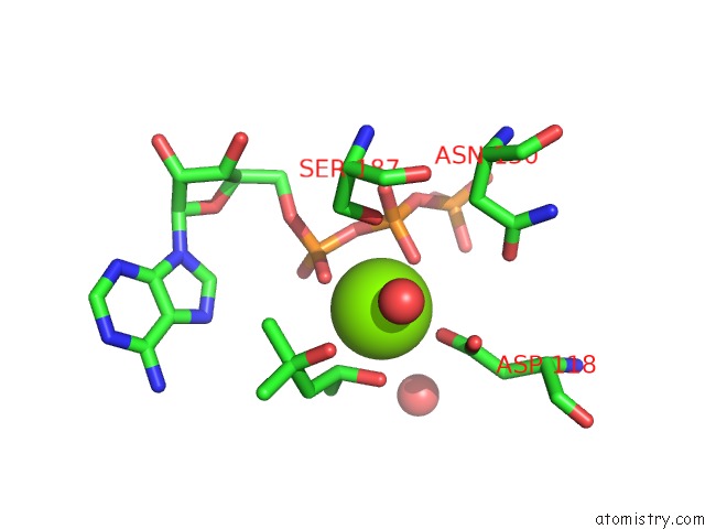
Mono view
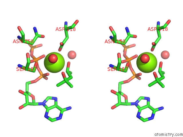
Stereo pair view
Mono view
Stereo pair view
A full contact list of Magnesium with other atoms in the Mg binding
site number 1 of Human Pyridoxal Kinase within 5.0Å range:
|
Magnesium binding site 2 out of 2 in 2yxu
Go back to
Magnesium binding site 2 out
of 2 in the Human Pyridoxal Kinase
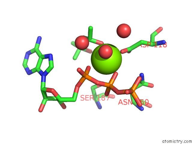
Mono view
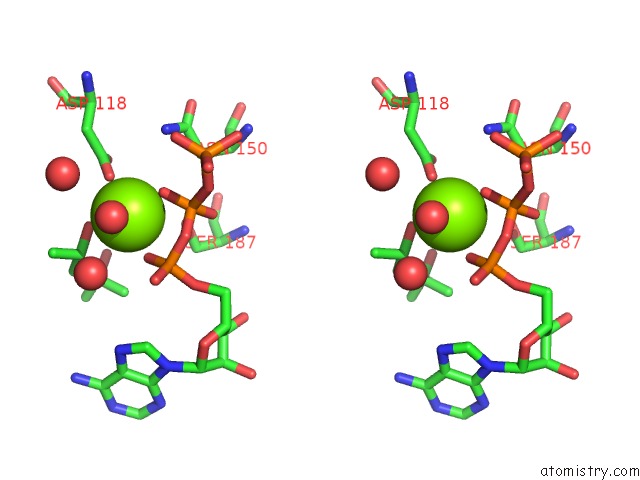
Stereo pair view
Mono view
Stereo pair view
A full contact list of Magnesium with other atoms in the Mg binding
site number 2 of Human Pyridoxal Kinase within 5.0Å range:
|
Reference:
F.N.Musayev,
M.L.Di Salvo,
T.P.Ko,
A.K.Gandhi,
A.Goswami,
V.Schirch,
M.K.Safo.
Crystal Structure of Human Pyridoxal Kinase: Structural Basis of M(+) and M(2+) Activation. Protein Sci. V. 16 2184 2007.
ISSN: ISSN 0961-8368
PubMed: 17766369
DOI: 10.1110/PS.073022107
Page generated: Sun Aug 10 16:49:44 2025
ISSN: ISSN 0961-8368
PubMed: 17766369
DOI: 10.1110/PS.073022107
Last articles
Mn in 9LJUMn in 9LJW
Mn in 9LJS
Mn in 9LJR
Mn in 9LJT
Mn in 9LJV
Mg in 9UA2
Mg in 9R96
Mg in 9VM1
Mg in 9P01