Magnesium »
PDB 3a0u-3abk »
3a28 »
Magnesium in PDB 3a28: Crystal Structure of L-2,3-Butanediol Dehydrogenase
Protein crystallography data
The structure of Crystal Structure of L-2,3-Butanediol Dehydrogenase, PDB code: 3a28
was solved by
M.Otagiri,
G.Kurisu,
S.Ui,
M.Kusunoki,
with X-Ray Crystallography technique. A brief refinement statistics is given in the table below:
| Resolution Low / High (Å) | 30.00 / 2.00 |
| Space group | P 1 |
| Cell size a, b, c (Å), α, β, γ (°) | 60.800, 69.200, 127.400, 96.10, 100.20, 109.60 |
| R / Rfree (%) | 19.3 / 24 |
Magnesium Binding Sites:
The binding sites of Magnesium atom in the Crystal Structure of L-2,3-Butanediol Dehydrogenase
(pdb code 3a28). This binding sites where shown within
5.0 Angstroms radius around Magnesium atom.
In total 4 binding sites of Magnesium where determined in the Crystal Structure of L-2,3-Butanediol Dehydrogenase, PDB code: 3a28:
Jump to Magnesium binding site number: 1; 2; 3; 4;
In total 4 binding sites of Magnesium where determined in the Crystal Structure of L-2,3-Butanediol Dehydrogenase, PDB code: 3a28:
Jump to Magnesium binding site number: 1; 2; 3; 4;
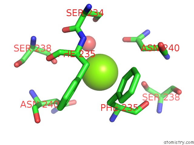
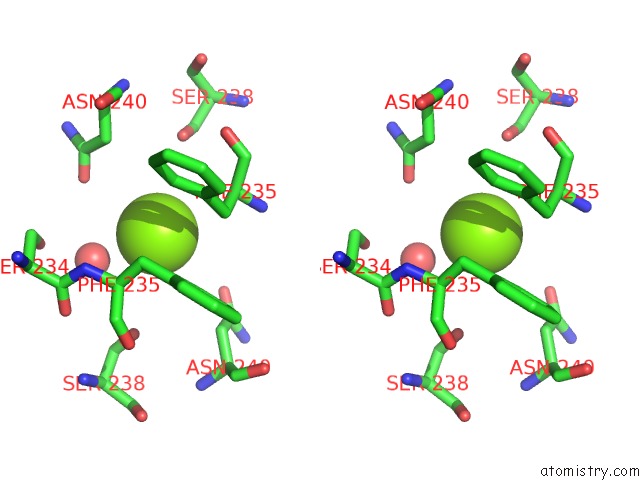
Magnesium binding site 1 out of 4 in 3a28
Go back to
Magnesium binding site 1 out
of 4 in the Crystal Structure of L-2,3-Butanediol Dehydrogenase
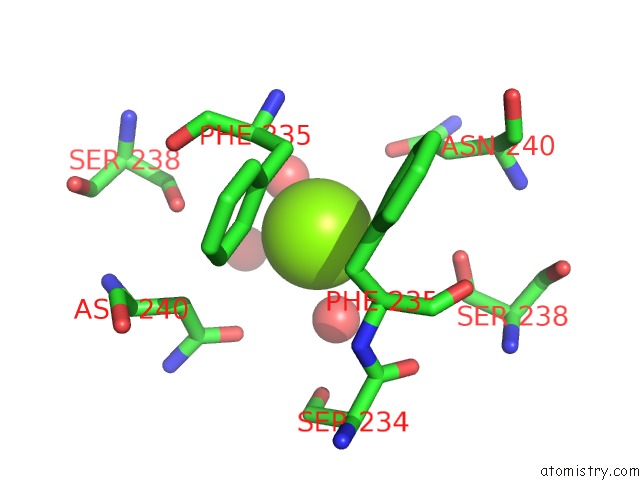
Mono view
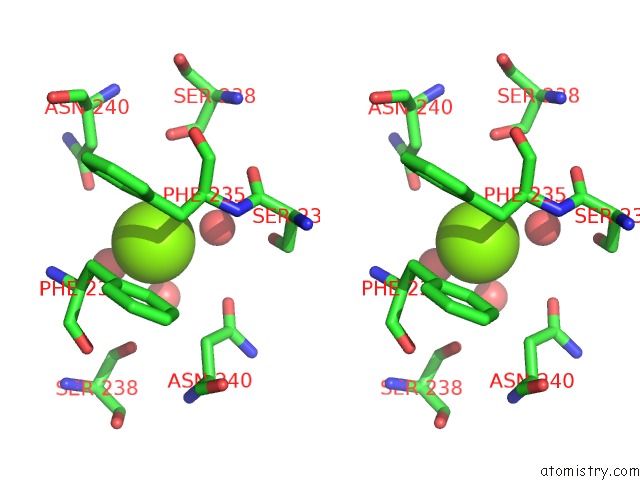
Stereo pair view
Mono view
Stereo pair view
A full contact list of Magnesium with other atoms in the Mg binding
site number 1 of Crystal Structure of L-2,3-Butanediol Dehydrogenase within 5.0Å range:
|
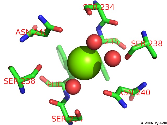
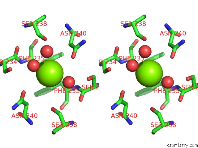
Magnesium binding site 2 out of 4 in 3a28
Go back to
Magnesium binding site 2 out
of 4 in the Crystal Structure of L-2,3-Butanediol Dehydrogenase
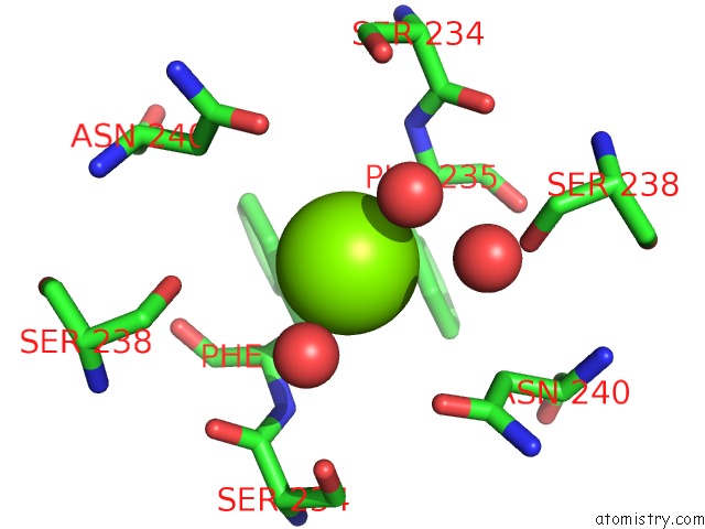
Mono view
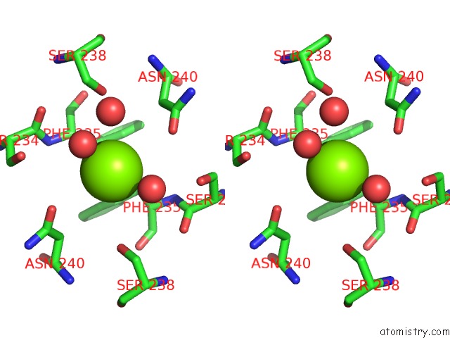
Stereo pair view
Mono view
Stereo pair view
A full contact list of Magnesium with other atoms in the Mg binding
site number 2 of Crystal Structure of L-2,3-Butanediol Dehydrogenase within 5.0Å range:
|
Magnesium binding site 3 out of 4 in 3a28
Go back to
Magnesium binding site 3 out
of 4 in the Crystal Structure of L-2,3-Butanediol Dehydrogenase
Mono view
Stereo pair view
Mono view
Stereo pair view
A full contact list of Magnesium with other atoms in the Mg binding
site number 3 of Crystal Structure of L-2,3-Butanediol Dehydrogenase within 5.0Å range:
|
Magnesium binding site 4 out of 4 in 3a28
Go back to
Magnesium binding site 4 out
of 4 in the Crystal Structure of L-2,3-Butanediol Dehydrogenase
Mono view
Stereo pair view
Mono view
Stereo pair view
A full contact list of Magnesium with other atoms in the Mg binding
site number 4 of Crystal Structure of L-2,3-Butanediol Dehydrogenase within 5.0Å range:
|
Reference:
M.Otagiri,
S.Ui,
Y.Takusagawa,
T.Ohtsuki,
G.Kurisu,
M.Kusunoki.
Structural Basis For Chiral Substrate Recognition By Two 2,3-Butanediol Dehydrogenases Febs Lett. V. 584 219 2010.
ISSN: ISSN 0014-5793
PubMed: 19941855
DOI: 10.1016/J.FEBSLET.2009.11.068
Page generated: Wed Aug 14 08:26:29 2024
ISSN: ISSN 0014-5793
PubMed: 19941855
DOI: 10.1016/J.FEBSLET.2009.11.068
Last articles
F in 8HSRF in 8HSL
F in 8HSJ
F in 8HUP
F in 8HUO
F in 8HUN
F in 8HUG
F in 8HTB
F in 8HOQ
F in 8HQ4