Magnesium »
PDB 3abl-3aln »
3ahg »
Magnesium in PDB 3ahg: H64A Mutant of Phosphoketolase From Bifidobacterium Breve Complexed with A Tricyclic Ring Form of Thiamine Diphosphate
Enzymatic activity of H64A Mutant of Phosphoketolase From Bifidobacterium Breve Complexed with A Tricyclic Ring Form of Thiamine Diphosphate
All present enzymatic activity of H64A Mutant of Phosphoketolase From Bifidobacterium Breve Complexed with A Tricyclic Ring Form of Thiamine Diphosphate:
4.1.2.22;
4.1.2.22;
Protein crystallography data
The structure of H64A Mutant of Phosphoketolase From Bifidobacterium Breve Complexed with A Tricyclic Ring Form of Thiamine Diphosphate, PDB code: 3ahg
was solved by
R.Suzuki,
T.Katayama,
B.-J.Kim,
T.Wakagi,
H.Shoun,
H.Ashida,
K.Yamamoto,
S.Fushinobu,
with X-Ray Crystallography technique. A brief refinement statistics is given in the table below:
| Resolution Low / High (Å) | 33.50 / 1.90 |
| Space group | I 4 2 2 |
| Cell size a, b, c (Å), α, β, γ (°) | 175.035, 175.035, 163.597, 90.00, 90.00, 90.00 |
| R / Rfree (%) | 16.3 / 20.5 |
Other elements in 3ahg:
The structure of H64A Mutant of Phosphoketolase From Bifidobacterium Breve Complexed with A Tricyclic Ring Form of Thiamine Diphosphate also contains other interesting chemical elements:
| Sodium | (Na) | 1 atom |
Magnesium Binding Sites:
The binding sites of Magnesium atom in the H64A Mutant of Phosphoketolase From Bifidobacterium Breve Complexed with A Tricyclic Ring Form of Thiamine Diphosphate
(pdb code 3ahg). This binding sites where shown within
5.0 Angstroms radius around Magnesium atom.
In total only one binding site of Magnesium was determined in the H64A Mutant of Phosphoketolase From Bifidobacterium Breve Complexed with A Tricyclic Ring Form of Thiamine Diphosphate, PDB code: 3ahg:
In total only one binding site of Magnesium was determined in the H64A Mutant of Phosphoketolase From Bifidobacterium Breve Complexed with A Tricyclic Ring Form of Thiamine Diphosphate, PDB code: 3ahg:
Magnesium binding site 1 out of 1 in 3ahg
Go back to
Magnesium binding site 1 out
of 1 in the H64A Mutant of Phosphoketolase From Bifidobacterium Breve Complexed with A Tricyclic Ring Form of Thiamine Diphosphate
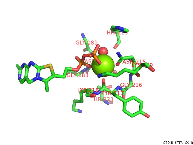
Mono view
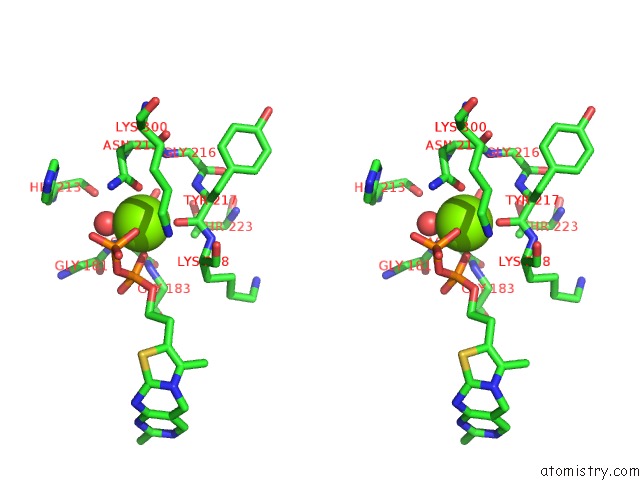
Stereo pair view
Mono view
Stereo pair view
A full contact list of Magnesium with other atoms in the Mg binding
site number 1 of H64A Mutant of Phosphoketolase From Bifidobacterium Breve Complexed with A Tricyclic Ring Form of Thiamine Diphosphate within 5.0Å range:
|
Reference:
R.Suzuki,
T.Katayama,
B.-J.Kim,
T.Wakagi,
H.Shoun,
H.Ashida,
K.Yamamoto,
S.Fushinobu.
Crystal Structures of Phosphoketolase: Thiamine Diphosphate-Dependent Dehydration Mechanism J.Biol.Chem. V. 285 34279 2010.
ISSN: ISSN 0021-9258
PubMed: 20739284
DOI: 10.1074/JBC.M110.156281
Page generated: Sun Aug 10 17:27:28 2025
ISSN: ISSN 0021-9258
PubMed: 20739284
DOI: 10.1074/JBC.M110.156281
Last articles
Mo in 2A9DMo in 2A9C
Mo in 2A9A
Mo in 2A9B
Mo in 2A99
Mo in 1ZXI
Mo in 1XDQ
Mo in 2A3P
Mo in 1Z13
Mo in 1Y5N