Magnesium »
PDB 3dfy-3dts »
3dk5 »
Magnesium in PDB 3dk5: Crystal Structure of Apo-Glmu From Mycobacterium Tuberculosis
Enzymatic activity of Crystal Structure of Apo-Glmu From Mycobacterium Tuberculosis
All present enzymatic activity of Crystal Structure of Apo-Glmu From Mycobacterium Tuberculosis:
2.3.1.157; 2.7.7.23;
2.3.1.157; 2.7.7.23;
Protein crystallography data
The structure of Crystal Structure of Apo-Glmu From Mycobacterium Tuberculosis, PDB code: 3dk5
was solved by
S.K.Verma,
B.Prakash,
with X-Ray Crystallography technique. A brief refinement statistics is given in the table below:
| Resolution Low / High (Å) | 29.30 / 2.23 |
| Space group | H 3 |
| Cell size a, b, c (Å), α, β, γ (°) | 79.600, 79.600, 278.000, 90.00, 90.00, 120.00 |
| R / Rfree (%) | 22.5 / 27.1 |
Magnesium Binding Sites:
The binding sites of Magnesium atom in the Crystal Structure of Apo-Glmu From Mycobacterium Tuberculosis
(pdb code 3dk5). This binding sites where shown within
5.0 Angstroms radius around Magnesium atom.
In total 2 binding sites of Magnesium where determined in the Crystal Structure of Apo-Glmu From Mycobacterium Tuberculosis, PDB code: 3dk5:
Jump to Magnesium binding site number: 1; 2;
In total 2 binding sites of Magnesium where determined in the Crystal Structure of Apo-Glmu From Mycobacterium Tuberculosis, PDB code: 3dk5:
Jump to Magnesium binding site number: 1; 2;
Magnesium binding site 1 out of 2 in 3dk5
Go back to
Magnesium binding site 1 out
of 2 in the Crystal Structure of Apo-Glmu From Mycobacterium Tuberculosis
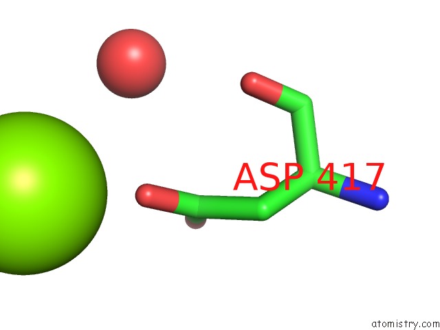
Mono view
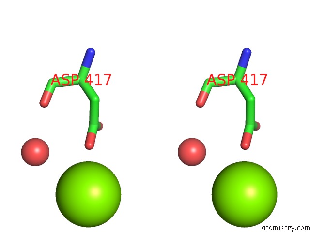
Stereo pair view
Mono view
Stereo pair view
A full contact list of Magnesium with other atoms in the Mg binding
site number 1 of Crystal Structure of Apo-Glmu From Mycobacterium Tuberculosis within 5.0Å range:
|
Magnesium binding site 2 out of 2 in 3dk5
Go back to
Magnesium binding site 2 out
of 2 in the Crystal Structure of Apo-Glmu From Mycobacterium Tuberculosis
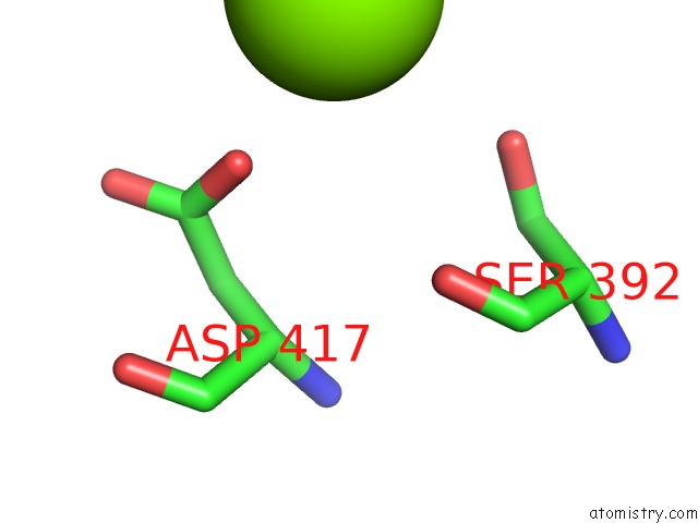
Mono view

Stereo pair view
Mono view
Stereo pair view
A full contact list of Magnesium with other atoms in the Mg binding
site number 2 of Crystal Structure of Apo-Glmu From Mycobacterium Tuberculosis within 5.0Å range:
|
Reference:
A.Parikh,
S.K.Verma,
S.Khan,
B.Prakash,
V.K.Nandicoori.
Pknb-Mediated Phosphorylation of A Novel Substrate, N-Acetylglucosamine-1-Phosphate Uridyltransferase, Modulates Its Acetyltransferase Activity. J.Mol.Biol. V. 386 451 2009.
ISSN: ISSN 0022-2836
PubMed: 19121323
DOI: 10.1016/J.JMB.2008.12.031
Page generated: Sun Aug 10 20:07:47 2025
ISSN: ISSN 0022-2836
PubMed: 19121323
DOI: 10.1016/J.JMB.2008.12.031
Last articles
Mg in 5QK2Mg in 5QK3
Mg in 5QK1
Mg in 5QK0
Mg in 5QJZ
Mg in 5QJY
Mg in 5QJX
Mg in 5QJW
Mg in 5QJV
Mg in 5QJU