Magnesium »
PDB 3dtu-3e35 »
3e25 »
Magnesium in PDB 3e25: Crystal Structure of M. Tuberculosis Glucosyl-3- Phosphoglycerate Synthase
Protein crystallography data
The structure of Crystal Structure of M. Tuberculosis Glucosyl-3- Phosphoglycerate Synthase, PDB code: 3e25
was solved by
P.J.B.Pereira,
N.Empadinhas,
M.S.Costa,
S.Macedo-Ribeiro,
with X-Ray Crystallography technique. A brief refinement statistics is given in the table below:
| Resolution Low / High (Å) | 32.34 / 2.70 |
| Space group | I 41 |
| Cell size a, b, c (Å), α, β, γ (°) | 100.309, 100.309, 127.035, 90.00, 90.00, 90.00 |
| R / Rfree (%) | 20.4 / 23.5 |
Magnesium Binding Sites:
The binding sites of Magnesium atom in the Crystal Structure of M. Tuberculosis Glucosyl-3- Phosphoglycerate Synthase
(pdb code 3e25). This binding sites where shown within
5.0 Angstroms radius around Magnesium atom.
In total only one binding site of Magnesium was determined in the Crystal Structure of M. Tuberculosis Glucosyl-3- Phosphoglycerate Synthase, PDB code: 3e25:
In total only one binding site of Magnesium was determined in the Crystal Structure of M. Tuberculosis Glucosyl-3- Phosphoglycerate Synthase, PDB code: 3e25:
Magnesium binding site 1 out of 1 in 3e25
Go back to
Magnesium binding site 1 out
of 1 in the Crystal Structure of M. Tuberculosis Glucosyl-3- Phosphoglycerate Synthase
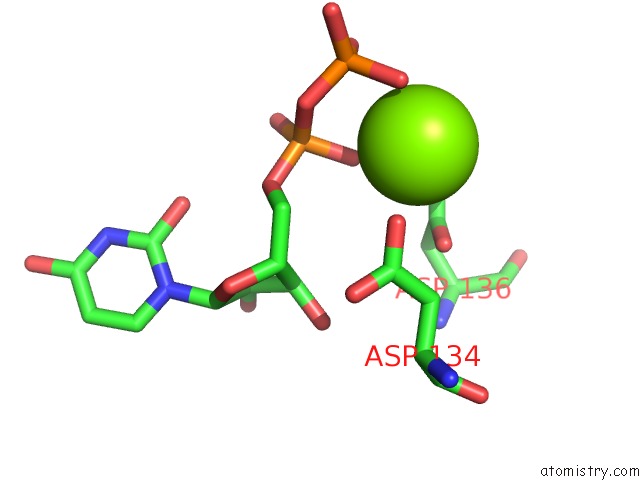
Mono view
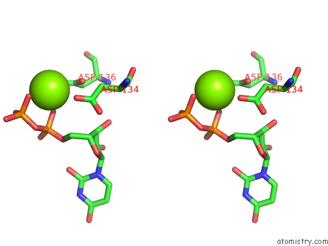
Stereo pair view
Mono view
Stereo pair view
A full contact list of Magnesium with other atoms in the Mg binding
site number 1 of Crystal Structure of M. Tuberculosis Glucosyl-3- Phosphoglycerate Synthase within 5.0Å range:
|
Reference:
P.J.B.Pereira,
N.Empadinhas,
L.Albuquerque,
B.Sa-Moura,
M.S.Da Costa,
S.Macedo-Ribeiro.
Mycobacterium Tuberculosis Glucosyl-3-Phosphoglycerate Synthase: Structure of A Key Enzyme in Methylglucose Lipopolysaccharide Biosynthesis Plos One V. 3 E3748 2008.
ISSN: ESSN 1932-6203
PubMed: 19015727
DOI: 10.1371/JOURNAL.PONE.0003748
Page generated: Wed Aug 14 12:50:13 2024
ISSN: ESSN 1932-6203
PubMed: 19015727
DOI: 10.1371/JOURNAL.PONE.0003748
Last articles
Fe in 5TISFe in 5TWT
Fe in 5TQN
Fe in 5TQO
Fe in 5TQP
Fe in 5TPG
Fe in 5TK5
Fe in 5TL8
Fe in 5T5I
Fe in 5TH5