Magnesium »
PDB 3dtu-3e35 »
3e2v »
Magnesium in PDB 3e2v: Crystal Structure of An Uncharacterized Amidohydrolase From Saccharomyces Cerevisiae
Protein crystallography data
The structure of Crystal Structure of An Uncharacterized Amidohydrolase From Saccharomyces Cerevisiae, PDB code: 3e2v
was solved by
J.B.Bonanno,
M.Dickey,
K.T.Bain,
S.Hu,
R.Romero,
D.Smith,
S.Wasserman,
J.M.Sauder,
S.K.Burley,
S.C.Almo,
New York Sgx Research Center Forstructural Genomics (Nysgxrc),
with X-Ray Crystallography technique. A brief refinement statistics is given in the table below:
| Resolution Low / High (Å) | 15.00 / 1.50 |
| Space group | P 1 21 1 |
| Cell size a, b, c (Å), α, β, γ (°) | 62.532, 55.007, 115.260, 90.00, 95.85, 90.00 |
| R / Rfree (%) | 17.3 / 19.9 |
Magnesium Binding Sites:
The binding sites of Magnesium atom in the Crystal Structure of An Uncharacterized Amidohydrolase From Saccharomyces Cerevisiae
(pdb code 3e2v). This binding sites where shown within
5.0 Angstroms radius around Magnesium atom.
In total 3 binding sites of Magnesium where determined in the Crystal Structure of An Uncharacterized Amidohydrolase From Saccharomyces Cerevisiae, PDB code: 3e2v:
Jump to Magnesium binding site number: 1; 2; 3;
In total 3 binding sites of Magnesium where determined in the Crystal Structure of An Uncharacterized Amidohydrolase From Saccharomyces Cerevisiae, PDB code: 3e2v:
Jump to Magnesium binding site number: 1; 2; 3;
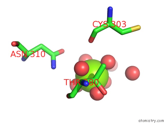
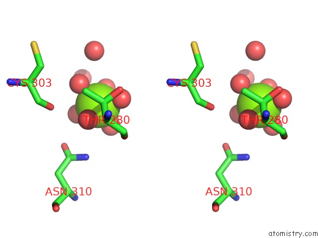
Magnesium binding site 1 out of 3 in 3e2v
Go back to
Magnesium binding site 1 out
of 3 in the Crystal Structure of An Uncharacterized Amidohydrolase From Saccharomyces Cerevisiae
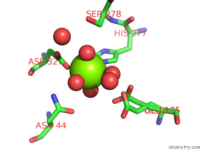
Mono view
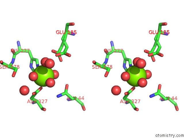
Stereo pair view
Mono view
Stereo pair view
A full contact list of Magnesium with other atoms in the Mg binding
site number 1 of Crystal Structure of An Uncharacterized Amidohydrolase From Saccharomyces Cerevisiae within 5.0Å range:
|
Magnesium binding site 2 out of 3 in 3e2v
Go back to
Magnesium binding site 2 out
of 3 in the Crystal Structure of An Uncharacterized Amidohydrolase From Saccharomyces Cerevisiae
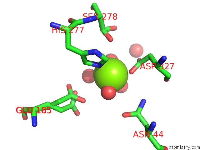
Mono view
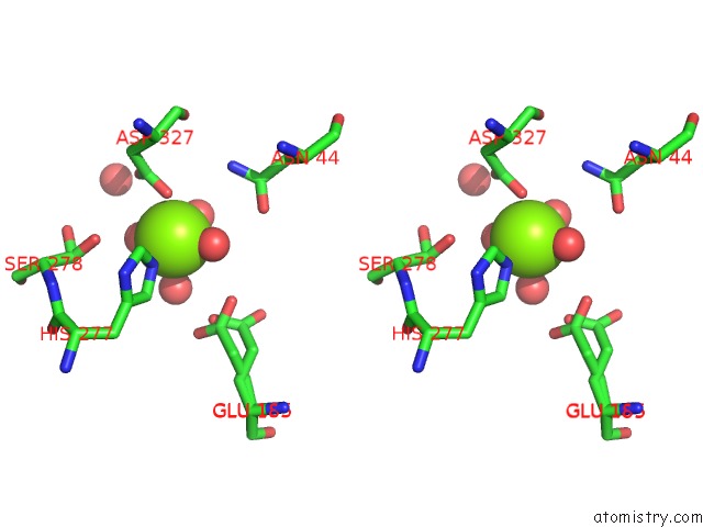
Stereo pair view
Mono view
Stereo pair view
A full contact list of Magnesium with other atoms in the Mg binding
site number 2 of Crystal Structure of An Uncharacterized Amidohydrolase From Saccharomyces Cerevisiae within 5.0Å range:
|
Magnesium binding site 3 out of 3 in 3e2v
Go back to
Magnesium binding site 3 out
of 3 in the Crystal Structure of An Uncharacterized Amidohydrolase From Saccharomyces Cerevisiae
Mono view
Stereo pair view
Mono view
Stereo pair view
A full contact list of Magnesium with other atoms in the Mg binding
site number 3 of Crystal Structure of An Uncharacterized Amidohydrolase From Saccharomyces Cerevisiae within 5.0Å range:
|
Reference:
J.B.Bonanno,
M.Dickey,
K.T.Bain,
S.Hu,
R.Romero,
D.Smith,
S.Wasserman,
J.M.Sauder,
S.K.Burley,
S.C.Almo.
Crystal Structure of An Uncharacterized Amidohydrolase From Saccharomyces Cerevisiae To Be Published.
Page generated: Wed Aug 14 12:51:59 2024
Last articles
Fe in 5TISFe in 5TWT
Fe in 5TQN
Fe in 5TQO
Fe in 5TQP
Fe in 5TPG
Fe in 5TK5
Fe in 5TL8
Fe in 5T5I
Fe in 5TH5