Magnesium »
PDB 3fcw-3fpk »
3fcx »
Magnesium in PDB 3fcx: Crystal Structure of Human Esterase D
Enzymatic activity of Crystal Structure of Human Esterase D
All present enzymatic activity of Crystal Structure of Human Esterase D:
3.1.2.12;
3.1.2.12;
Protein crystallography data
The structure of Crystal Structure of Human Esterase D, PDB code: 3fcx
was solved by
D.Wu,
Y.Li,
G.Song,
D.Zhang,
N.Shaw,
Z.J.Liu,
with X-Ray Crystallography technique. A brief refinement statistics is given in the table below:
| Resolution Low / High (Å) | 40.16 / 1.50 |
| Space group | P 1 21 1 |
| Cell size a, b, c (Å), α, β, γ (°) | 51.542, 70.724, 65.014, 90.00, 108.84, 90.00 |
| R / Rfree (%) | 17.6 / 19.6 |
Other elements in 3fcx:
The structure of Crystal Structure of Human Esterase D also contains other interesting chemical elements:
| Calcium | (Ca) | 3 atoms |
Magnesium Binding Sites:
The binding sites of Magnesium atom in the Crystal Structure of Human Esterase D
(pdb code 3fcx). This binding sites where shown within
5.0 Angstroms radius around Magnesium atom.
In total only one binding site of Magnesium was determined in the Crystal Structure of Human Esterase D, PDB code: 3fcx:
In total only one binding site of Magnesium was determined in the Crystal Structure of Human Esterase D, PDB code: 3fcx:
Magnesium binding site 1 out of 1 in 3fcx
Go back to
Magnesium binding site 1 out
of 1 in the Crystal Structure of Human Esterase D
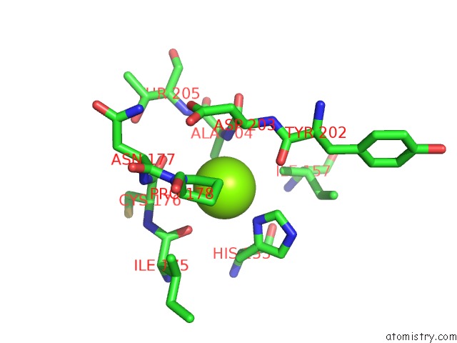
Mono view
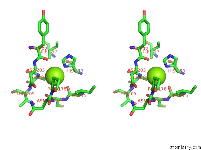
Stereo pair view
Mono view
Stereo pair view
A full contact list of Magnesium with other atoms in the Mg binding
site number 1 of Crystal Structure of Human Esterase D within 5.0Å range:
|
Reference:
D.Wu,
Y.Li,
G.Song,
D.Zhang,
N.Shaw,
Z.J.Liu.
Crystal Structure of Human Esterase D: A Potential Genetic Marker of Retinoblastoma Faseb J. V. 23 1441 2009.
ISSN: ISSN 0892-6638
PubMed: 19126594
DOI: 10.1096/FJ.08-125286
Page generated: Wed Aug 14 13:43:26 2024
ISSN: ISSN 0892-6638
PubMed: 19126594
DOI: 10.1096/FJ.08-125286
Last articles
Mg in 3ATTMg in 3ATS
Mg in 3ARN
Mg in 3ATU
Mg in 3ASO
Mg in 3ATF
Mg in 3ASN
Mg in 3AT9
Mg in 3ARA
Mg in 3AS5