Magnesium »
PDB 3fcw-3fpk »
3ffa »
Magnesium in PDB 3ffa: Crystal Structure of A Fast Activating G Protein Mutant
Protein crystallography data
The structure of Crystal Structure of A Fast Activating G Protein Mutant, PDB code: 3ffa
was solved by
R.Chauhan,
N.Kapoor,
with X-Ray Crystallography technique. A brief refinement statistics is given in the table below:
| Resolution Low / High (Å) | 57.40 / 2.30 |
| Space group | P 32 2 1 |
| Cell size a, b, c (Å), α, β, γ (°) | 79.008, 79.008, 105.667, 90.00, 90.00, 120.00 |
| R / Rfree (%) | 18.5 / 25.2 |
Magnesium Binding Sites:
The binding sites of Magnesium atom in the Crystal Structure of A Fast Activating G Protein Mutant
(pdb code 3ffa). This binding sites where shown within
5.0 Angstroms radius around Magnesium atom.
In total only one binding site of Magnesium was determined in the Crystal Structure of A Fast Activating G Protein Mutant, PDB code: 3ffa:
In total only one binding site of Magnesium was determined in the Crystal Structure of A Fast Activating G Protein Mutant, PDB code: 3ffa:
Magnesium binding site 1 out of 1 in 3ffa
Go back to
Magnesium binding site 1 out
of 1 in the Crystal Structure of A Fast Activating G Protein Mutant
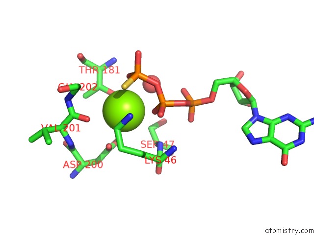
Mono view
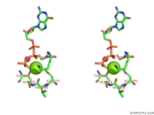
Stereo pair view
Mono view
Stereo pair view
A full contact list of Magnesium with other atoms in the Mg binding
site number 1 of Crystal Structure of A Fast Activating G Protein Mutant within 5.0Å range:
|
Reference:
N.Kapoor,
S.T.Menon,
R.Chauhan,
P.Sachdev,
T.P.Sakmar.
Structural Evidence For A Sequential Release Mechanism For Activation of Heterotrimeric G Proteins. J.Mol.Biol. V. 393 882 2009.
ISSN: ISSN 0022-2836
PubMed: 19703466
DOI: 10.1016/J.JMB.2009.08.043
Page generated: Sun Aug 10 20:58:52 2025
ISSN: ISSN 0022-2836
PubMed: 19703466
DOI: 10.1016/J.JMB.2009.08.043
Last articles
Mg in 4DUYMg in 4DR7
Mg in 4DR6
Mg in 4DR5
Mg in 4DUX
Mg in 4DUW
Mg in 4DUV
Mg in 4DUO
Mg in 4DUG
Mg in 4DTY