Magnesium »
PDB 3fcw-3fpk »
3fhd »
Magnesium in PDB 3fhd: Crystal Structure of the Shutoff and Exonuclease Protein From Kaposis Sarcoma Associated Herpesvirus
Protein crystallography data
The structure of Crystal Structure of the Shutoff and Exonuclease Protein From Kaposis Sarcoma Associated Herpesvirus, PDB code: 3fhd
was solved by
S.L.Dahlroth,
D.Gurmu,
F.Schmitzberger,
J.Haas,
H.Erlandsen,
P.Nordlund,
with X-Ray Crystallography technique. A brief refinement statistics is given in the table below:
| Resolution Low / High (Å) | 46.63 / 1.85 |
| Space group | P 1 |
| Cell size a, b, c (Å), α, β, γ (°) | 43.247, 48.905, 67.294, 95.48, 106.86, 104.27 |
| R / Rfree (%) | 18.4 / 22.8 |
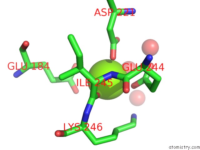
Magnesium Binding Sites:
The binding sites of Magnesium atom in the Crystal Structure of the Shutoff and Exonuclease Protein From Kaposis Sarcoma Associated Herpesvirus
(pdb code 3fhd). This binding sites where shown within
5.0 Angstroms radius around Magnesium atom.
In total only one binding site of Magnesium was determined in the Crystal Structure of the Shutoff and Exonuclease Protein From Kaposis Sarcoma Associated Herpesvirus, PDB code: 3fhd:
In total only one binding site of Magnesium was determined in the Crystal Structure of the Shutoff and Exonuclease Protein From Kaposis Sarcoma Associated Herpesvirus, PDB code: 3fhd:
Magnesium binding site 1 out of 1 in 3fhd
Go back to
Magnesium binding site 1 out
of 1 in the Crystal Structure of the Shutoff and Exonuclease Protein From Kaposis Sarcoma Associated Herpesvirus
Mono view
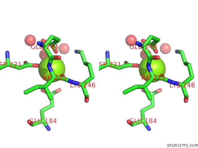
Stereo pair view
Mono view
Stereo pair view
A full contact list of Magnesium with other atoms in the Mg binding
site number 1 of Crystal Structure of the Shutoff and Exonuclease Protein From Kaposis Sarcoma Associated Herpesvirus within 5.0Å range:
|
Reference:
S.L.Dahlroth,
D.Gurmu,
F.Schmitzberger,
H.Engman,
J.Haas,
H.Erlandsen,
P.Nordlund.
Crystal Structure of the Shutoff and Exonuclease Protein From the Oncogenic Kaposi'S Sarcoma-Associated Herpesvirus Febs J. V. 276 6636 2009.
ISSN: ISSN 1742-464X
PubMed: 19843164
DOI: 10.1111/J.1742-4658.2009.07374.X
Page generated: Sun Aug 10 20:59:56 2025
ISSN: ISSN 1742-464X
PubMed: 19843164
DOI: 10.1111/J.1742-4658.2009.07374.X
Last articles
Mg in 3SDKMg in 3SDV
Mg in 3SDU
Mg in 3SDT
Mg in 3SDR
Mg in 3SBF
Mg in 3SB5
Mg in 3SCY
Mg in 3SBE
Mg in 3SBD