Magnesium »
PDB 3fps-3fzi »
3fyi »
Magnesium in PDB 3fyi: Catalytic Core Subunits (I and II) of Cytochrome C Oxidase From Rhodobacter Sphaeroides in the Reduced State Bound with Cyanide
Enzymatic activity of Catalytic Core Subunits (I and II) of Cytochrome C Oxidase From Rhodobacter Sphaeroides in the Reduced State Bound with Cyanide
All present enzymatic activity of Catalytic Core Subunits (I and II) of Cytochrome C Oxidase From Rhodobacter Sphaeroides in the Reduced State Bound with Cyanide:
1.9.3.1;
1.9.3.1;
Protein crystallography data
The structure of Catalytic Core Subunits (I and II) of Cytochrome C Oxidase From Rhodobacter Sphaeroides in the Reduced State Bound with Cyanide, PDB code: 3fyi
was solved by
L.Qin,
D.A.Mills,
D.A.Proshlyakov,
C.Hiser,
S.Ferguson-Miller,
with X-Ray Crystallography technique. A brief refinement statistics is given in the table below:
| Resolution Low / High (Å) | 50.00 / 2.20 |
| Space group | P 21 21 21 |
| Cell size a, b, c (Å), α, β, γ (°) | 124.344, 131.877, 176.160, 90.00, 90.00, 90.00 |
| R / Rfree (%) | 19.4 / 21.9 |
Other elements in 3fyi:
The structure of Catalytic Core Subunits (I and II) of Cytochrome C Oxidase From Rhodobacter Sphaeroides in the Reduced State Bound with Cyanide also contains other interesting chemical elements:
| Cadmium | (Cd) | 4 atoms |
| Iron | (Fe) | 4 atoms |
| Calcium | (Ca) | 2 atoms |
| Copper | (Cu) | 6 atoms |
Magnesium Binding Sites:
The binding sites of Magnesium atom in the Catalytic Core Subunits (I and II) of Cytochrome C Oxidase From Rhodobacter Sphaeroides in the Reduced State Bound with Cyanide
(pdb code 3fyi). This binding sites where shown within
5.0 Angstroms radius around Magnesium atom.
In total 2 binding sites of Magnesium where determined in the Catalytic Core Subunits (I and II) of Cytochrome C Oxidase From Rhodobacter Sphaeroides in the Reduced State Bound with Cyanide, PDB code: 3fyi:
Jump to Magnesium binding site number: 1; 2;
In total 2 binding sites of Magnesium where determined in the Catalytic Core Subunits (I and II) of Cytochrome C Oxidase From Rhodobacter Sphaeroides in the Reduced State Bound with Cyanide, PDB code: 3fyi:
Jump to Magnesium binding site number: 1; 2;
Magnesium binding site 1 out of 2 in 3fyi
Go back to
Magnesium binding site 1 out
of 2 in the Catalytic Core Subunits (I and II) of Cytochrome C Oxidase From Rhodobacter Sphaeroides in the Reduced State Bound with Cyanide
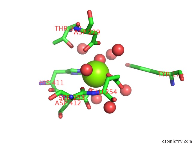
Mono view
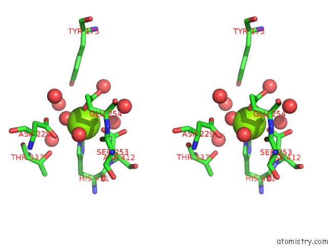
Stereo pair view
Mono view
Stereo pair view
A full contact list of Magnesium with other atoms in the Mg binding
site number 1 of Catalytic Core Subunits (I and II) of Cytochrome C Oxidase From Rhodobacter Sphaeroides in the Reduced State Bound with Cyanide within 5.0Å range:
|
Magnesium binding site 2 out of 2 in 3fyi
Go back to
Magnesium binding site 2 out
of 2 in the Catalytic Core Subunits (I and II) of Cytochrome C Oxidase From Rhodobacter Sphaeroides in the Reduced State Bound with Cyanide
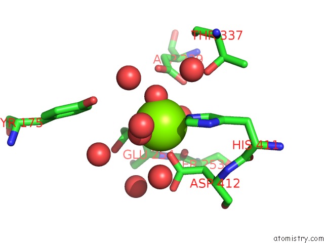
Mono view
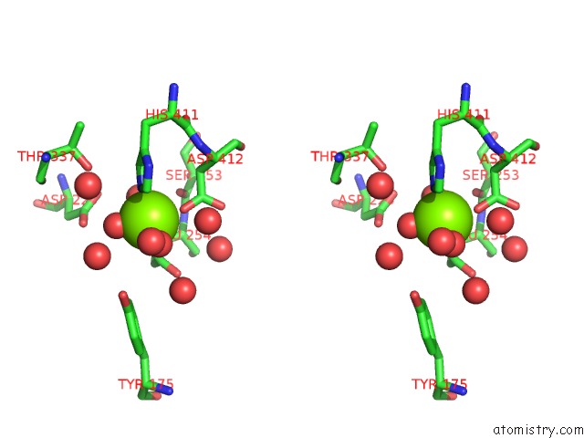
Stereo pair view
Mono view
Stereo pair view
A full contact list of Magnesium with other atoms in the Mg binding
site number 2 of Catalytic Core Subunits (I and II) of Cytochrome C Oxidase From Rhodobacter Sphaeroides in the Reduced State Bound with Cyanide within 5.0Å range:
|
Reference:
L.Qin,
J.Liu,
D.Mills,
D.A.Proshlyakov,
C.Hiser,
S.Ferguson-Miller.
Redox Dependent Conformational Changes in Cytochrome C Oxidase Suggest A Gating Mechanism For Proton Uptake. Biochemistry V. 48 5121 2009.
ISSN: ISSN 0006-2960
PubMed: 19397279
DOI: 10.1021/BI9001387
Page generated: Sun Aug 10 21:07:42 2025
ISSN: ISSN 0006-2960
PubMed: 19397279
DOI: 10.1021/BI9001387
Last articles
Na in 6UAONa in 6UAQ
Na in 6U8T
Na in 6U7P
Na in 6U9J
Na in 6U98
Na in 6U7O
Na in 6U6B
Na in 6U6U
Na in 6U6C