Magnesium »
PDB 3g8d-3gn6 »
3gao »
Magnesium in PDB 3gao: Crystal Structure of the Guanine Riboswitch Bound to Xanthine.
Protein crystallography data
The structure of Crystal Structure of the Guanine Riboswitch Bound to Xanthine., PDB code: 3gao
was solved by
S.D.Gilbert,
R.T.Batey,
with X-Ray Crystallography technique. A brief refinement statistics is given in the table below:
| Resolution Low / High (Å) | 19.49 / 1.90 |
| Space group | C 1 2 1 |
| Cell size a, b, c (Å), α, β, γ (°) | 131.351, 35.099, 42.248, 90.00, 90.38, 90.00 |
| R / Rfree (%) | 21 / 24.6 |
Other elements in 3gao:
The structure of Crystal Structure of the Guanine Riboswitch Bound to Xanthine. also contains other interesting chemical elements:
| Cobalt | (Co) | 13 atoms |
Magnesium Binding Sites:
The binding sites of Magnesium atom in the Crystal Structure of the Guanine Riboswitch Bound to Xanthine.
(pdb code 3gao). This binding sites where shown within
5.0 Angstroms radius around Magnesium atom.
In total only one binding site of Magnesium was determined in the Crystal Structure of the Guanine Riboswitch Bound to Xanthine., PDB code: 3gao:
In total only one binding site of Magnesium was determined in the Crystal Structure of the Guanine Riboswitch Bound to Xanthine., PDB code: 3gao:
Magnesium binding site 1 out of 1 in 3gao
Go back to
Magnesium binding site 1 out
of 1 in the Crystal Structure of the Guanine Riboswitch Bound to Xanthine.
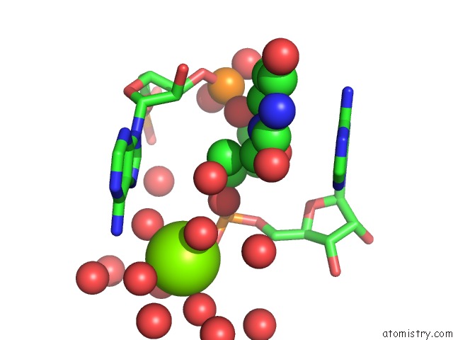
Mono view
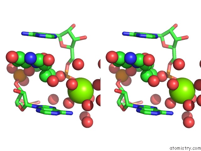
Stereo pair view
Mono view
Stereo pair view
A full contact list of Magnesium with other atoms in the Mg binding
site number 1 of Crystal Structure of the Guanine Riboswitch Bound to Xanthine. within 5.0Å range:
|
Reference:
S.D.Gilbert,
F.E.Reyes,
A.L.Edwards,
R.T.Batey.
Adaptive Ligand Binding By the Purine Riboswitch in the Recognition of Guanine and Adenine Analogs. Structure V. 17 857 2009.
ISSN: ISSN 0969-2126
PubMed: 19523903
DOI: 10.1016/J.STR.2009.04.009
Page generated: Sun Aug 10 21:39:11 2025
ISSN: ISSN 0969-2126
PubMed: 19523903
DOI: 10.1016/J.STR.2009.04.009
Last articles
Mg in 3NX3Mg in 3NYR
Mg in 3NYQ
Mg in 3NWN
Mg in 3NWO
Mg in 3NWE
Mg in 3NVS
Mg in 3NWB
Mg in 3NW9
Mg in 3NUH