Magnesium »
PDB 3g8d-3gn6 »
3gli »
Magnesium in PDB 3gli: Crystal Structure of the E. Coli Clamp Loader Bound to Primer-Template Dna and Psi Peptide
Enzymatic activity of Crystal Structure of the E. Coli Clamp Loader Bound to Primer-Template Dna and Psi Peptide
All present enzymatic activity of Crystal Structure of the E. Coli Clamp Loader Bound to Primer-Template Dna and Psi Peptide:
2.7.7.7;
2.7.7.7;
Protein crystallography data
The structure of Crystal Structure of the E. Coli Clamp Loader Bound to Primer-Template Dna and Psi Peptide, PDB code: 3gli
was solved by
K.R.Simonetta,
A.J.Cantor,
J.Kuriyan,
with X-Ray Crystallography technique. A brief refinement statistics is given in the table below:
| Resolution Low / High (Å) | 73.03 / 3.50 |
| Space group | P 21 21 21 |
| Cell size a, b, c (Å), α, β, γ (°) | 98.690, 217.180, 275.320, 90.00, 90.00, 90.00 |
| R / Rfree (%) | 22.2 / 25.7 |
Other elements in 3gli:
The structure of Crystal Structure of the E. Coli Clamp Loader Bound to Primer-Template Dna and Psi Peptide also contains other interesting chemical elements:
| Fluorine | (F) | 18 atoms |
| Zinc | (Zn) | 8 atoms |
Magnesium Binding Sites:
The binding sites of Magnesium atom in the Crystal Structure of the E. Coli Clamp Loader Bound to Primer-Template Dna and Psi Peptide
(pdb code 3gli). This binding sites where shown within
5.0 Angstroms radius around Magnesium atom.
In total 6 binding sites of Magnesium where determined in the Crystal Structure of the E. Coli Clamp Loader Bound to Primer-Template Dna and Psi Peptide, PDB code: 3gli:
Jump to Magnesium binding site number: 1; 2; 3; 4; 5; 6;
In total 6 binding sites of Magnesium where determined in the Crystal Structure of the E. Coli Clamp Loader Bound to Primer-Template Dna and Psi Peptide, PDB code: 3gli:
Jump to Magnesium binding site number: 1; 2; 3; 4; 5; 6;
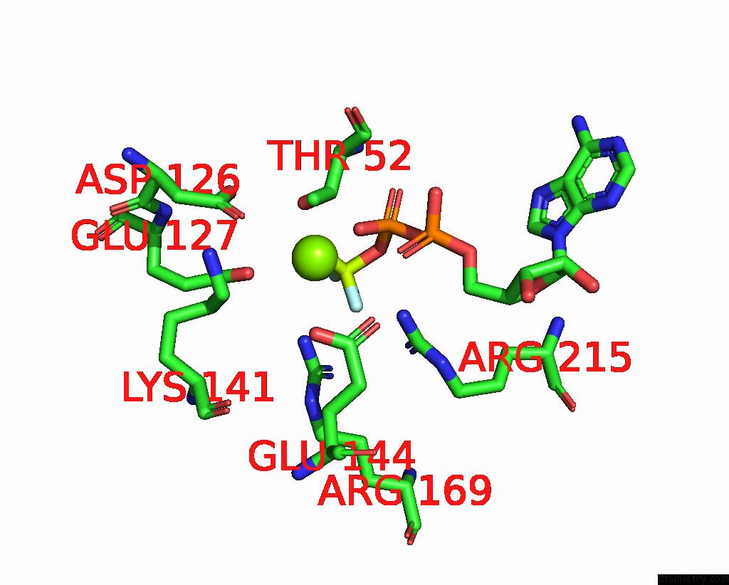
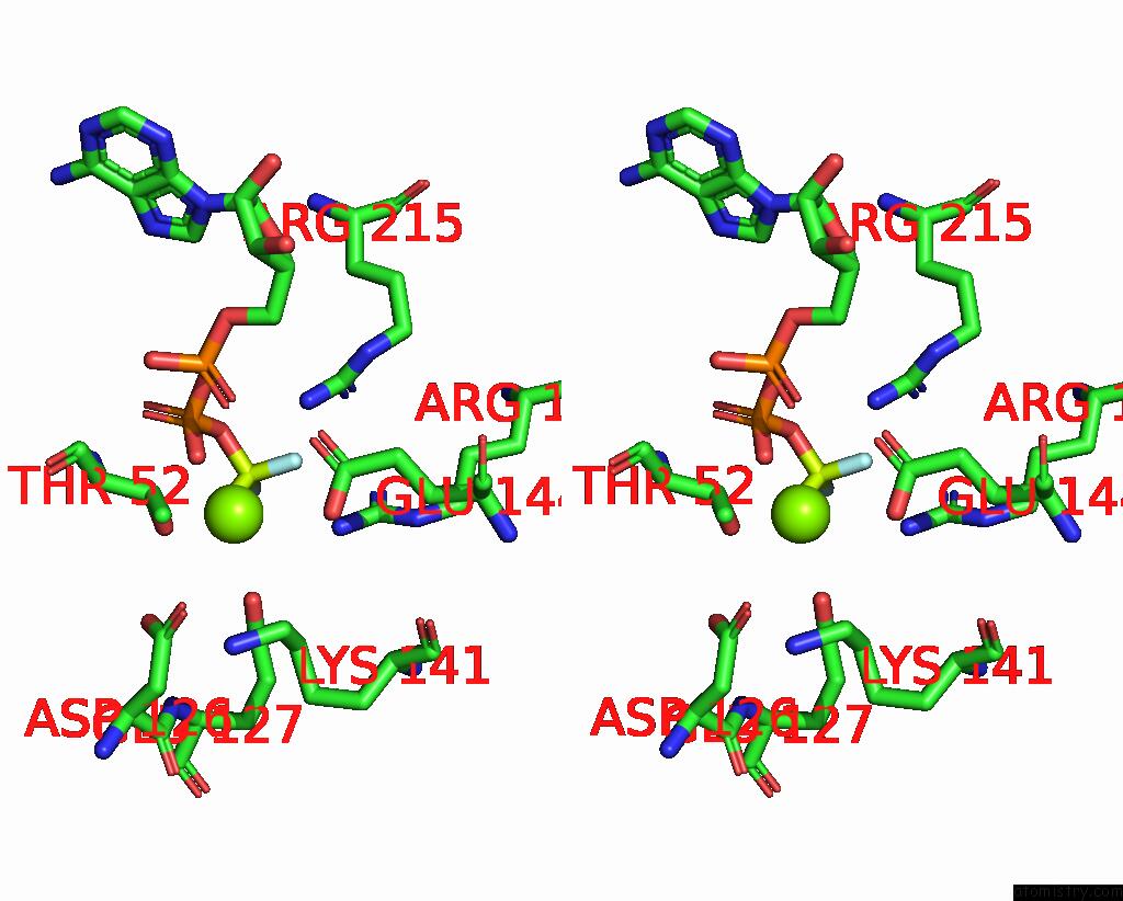
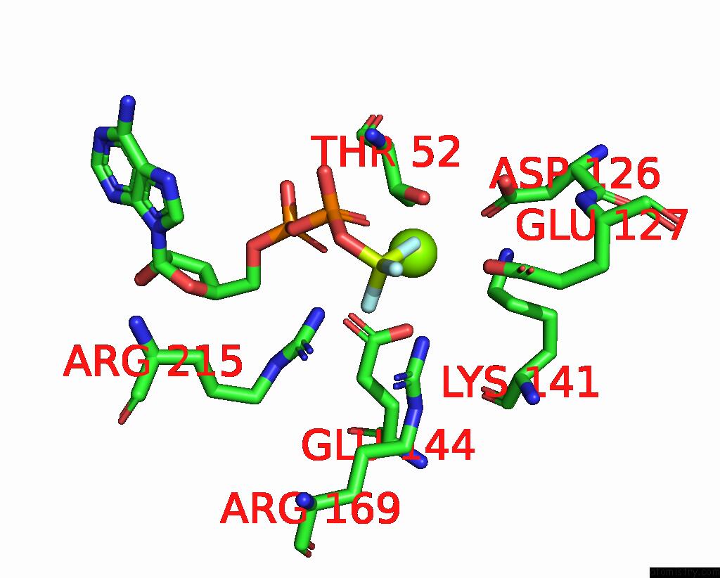
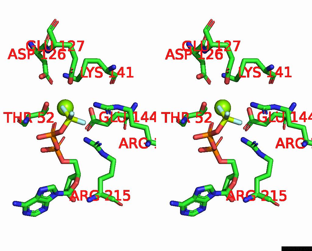
Magnesium binding site 1 out of 6 in 3gli
Go back to
Magnesium binding site 1 out
of 6 in the Crystal Structure of the E. Coli Clamp Loader Bound to Primer-Template Dna and Psi Peptide
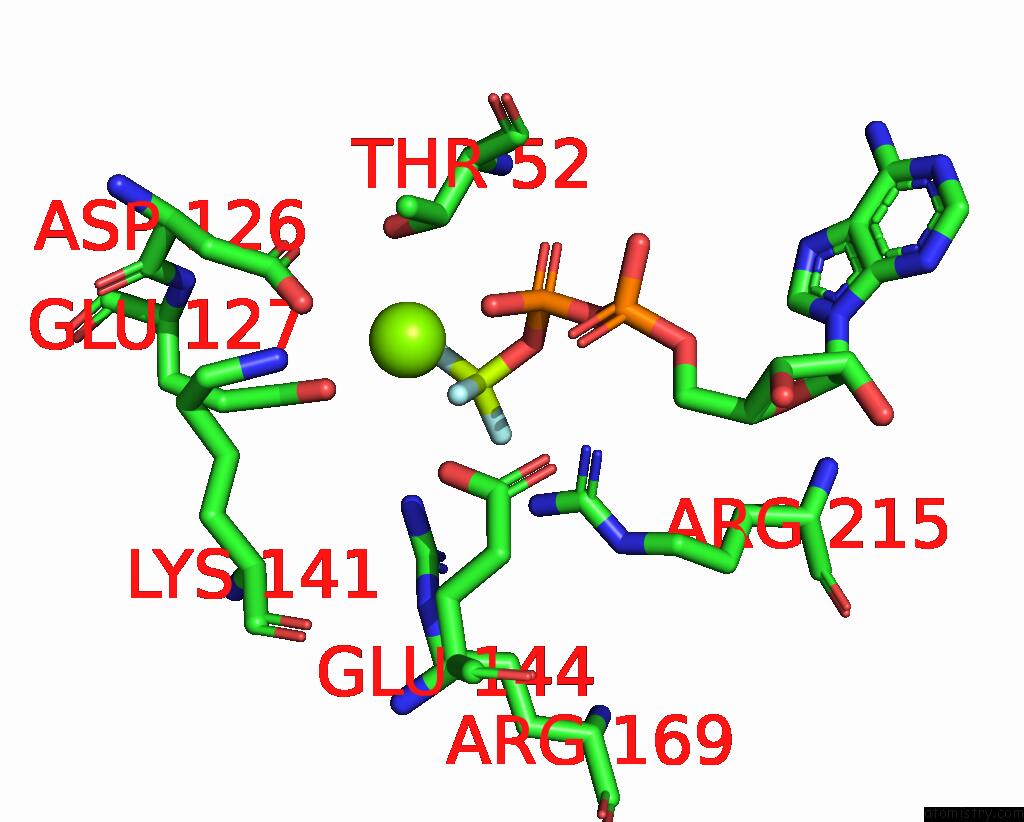
Mono view

Stereo pair view
Mono view
Stereo pair view
A full contact list of Magnesium with other atoms in the Mg binding
site number 1 of Crystal Structure of the E. Coli Clamp Loader Bound to Primer-Template Dna and Psi Peptide within 5.0Å range:
|
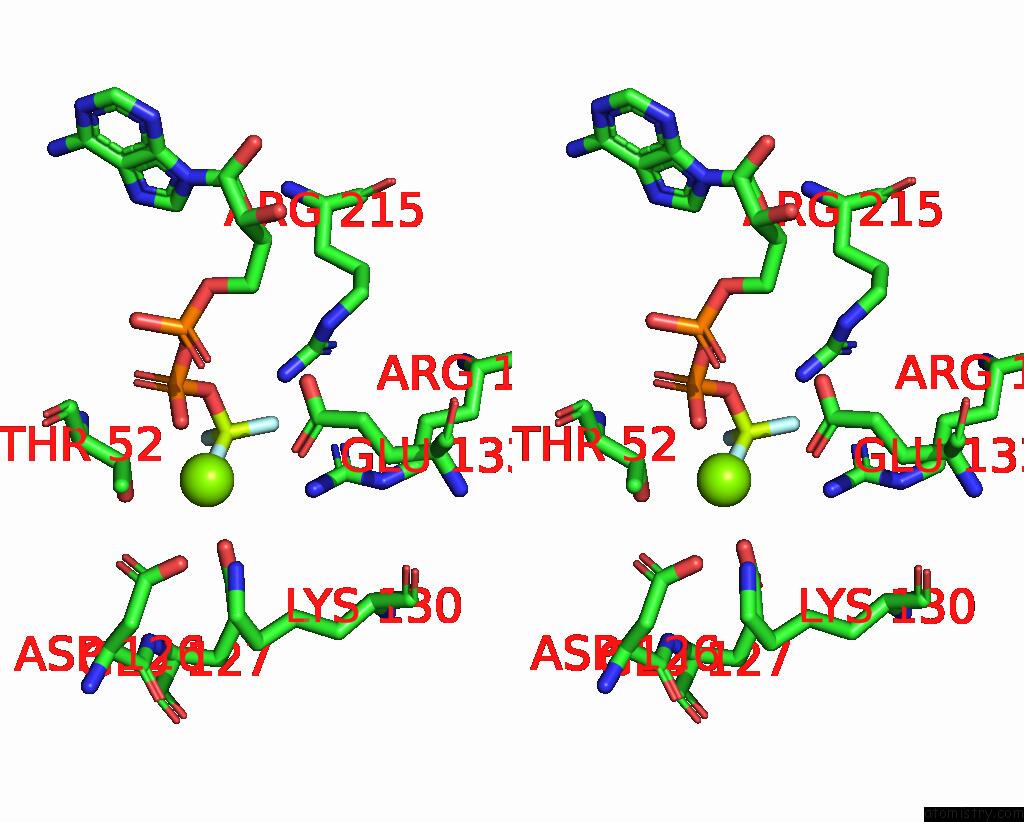
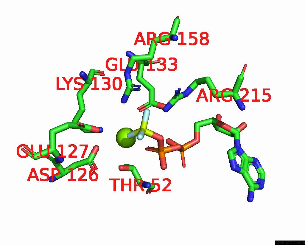
Magnesium binding site 2 out of 6 in 3gli
Go back to
Magnesium binding site 2 out
of 6 in the Crystal Structure of the E. Coli Clamp Loader Bound to Primer-Template Dna and Psi Peptide
Mono view
Stereo pair view
Mono view
Stereo pair view
A full contact list of Magnesium with other atoms in the Mg binding
site number 2 of Crystal Structure of the E. Coli Clamp Loader Bound to Primer-Template Dna and Psi Peptide within 5.0Å range:
|
Magnesium binding site 3 out of 6 in 3gli
Go back to
Magnesium binding site 3 out
of 6 in the Crystal Structure of the E. Coli Clamp Loader Bound to Primer-Template Dna and Psi Peptide

Mono view
Stereo pair view
Mono view
Stereo pair view
A full contact list of Magnesium with other atoms in the Mg binding
site number 3 of Crystal Structure of the E. Coli Clamp Loader Bound to Primer-Template Dna and Psi Peptide within 5.0Å range:
|
Magnesium binding site 4 out of 6 in 3gli
Go back to
Magnesium binding site 4 out
of 6 in the Crystal Structure of the E. Coli Clamp Loader Bound to Primer-Template Dna and Psi Peptide

Mono view

Stereo pair view
Mono view
Stereo pair view
A full contact list of Magnesium with other atoms in the Mg binding
site number 4 of Crystal Structure of the E. Coli Clamp Loader Bound to Primer-Template Dna and Psi Peptide within 5.0Å range:
|
Magnesium binding site 5 out of 6 in 3gli
Go back to
Magnesium binding site 5 out
of 6 in the Crystal Structure of the E. Coli Clamp Loader Bound to Primer-Template Dna and Psi Peptide
Mono view
Stereo pair view
Mono view
Stereo pair view
A full contact list of Magnesium with other atoms in the Mg binding
site number 5 of Crystal Structure of the E. Coli Clamp Loader Bound to Primer-Template Dna and Psi Peptide within 5.0Å range:
|
Magnesium binding site 6 out of 6 in 3gli
Go back to
Magnesium binding site 6 out
of 6 in the Crystal Structure of the E. Coli Clamp Loader Bound to Primer-Template Dna and Psi Peptide
Mono view

Stereo pair view
Mono view
Stereo pair view
A full contact list of Magnesium with other atoms in the Mg binding
site number 6 of Crystal Structure of the E. Coli Clamp Loader Bound to Primer-Template Dna and Psi Peptide within 5.0Å range:
|
Reference:
K.R.Simonetta,
S.L.Kazmirski,
E.R.Goedken,
A.J.Cantor,
B.A.Kelch,
R.Mcnally,
S.N.Seyedin,
D.L.Makino,
M.O'donnell,
J.Kuriyan.
The Mechanism of Atp-Dependent Primer-Template Recognition By A Clamp Loader Complex. Cell(Cambridge,Mass.) V. 137 659 2009.
ISSN: ISSN 0092-8674
PubMed: 19450514
DOI: 10.1016/J.CELL.2009.03.044
Page generated: Sun Aug 10 21:43:08 2025
ISSN: ISSN 0092-8674
PubMed: 19450514
DOI: 10.1016/J.CELL.2009.03.044
Last articles
Mg in 6TRAMg in 6TR4
Mg in 6TR3
Mg in 6TMF
Mg in 6TQO
Mg in 6TQN
Mg in 6TQF
Mg in 6TQE
Mg in 6TQB
Mg in 6TQA