Magnesium »
PDB 3gx3-3hd1 »
3h1f »
Magnesium in PDB 3h1f: Crystal Structure of Chey Mutant D53A of Helicobacter Pylori
Protein crystallography data
The structure of Crystal Structure of Chey Mutant D53A of Helicobacter Pylori, PDB code: 3h1f
was solved by
K.H.Lam,
T.K.Ling,
S.W.Au,
with X-Ray Crystallography technique. A brief refinement statistics is given in the table below:
| Resolution Low / High (Å) | 15.53 / 2.20 |
| Space group | C 1 2 1 |
| Cell size a, b, c (Å), α, β, γ (°) | 70.335, 38.081, 38.639, 90.00, 107.35, 90.00 |
| R / Rfree (%) | 18 / 23.3 |
Magnesium Binding Sites:
The binding sites of Magnesium atom in the Crystal Structure of Chey Mutant D53A of Helicobacter Pylori
(pdb code 3h1f). This binding sites where shown within
5.0 Angstroms radius around Magnesium atom.
In total only one binding site of Magnesium was determined in the Crystal Structure of Chey Mutant D53A of Helicobacter Pylori, PDB code: 3h1f:
In total only one binding site of Magnesium was determined in the Crystal Structure of Chey Mutant D53A of Helicobacter Pylori, PDB code: 3h1f:
Magnesium binding site 1 out of 1 in 3h1f
Go back to
Magnesium binding site 1 out
of 1 in the Crystal Structure of Chey Mutant D53A of Helicobacter Pylori
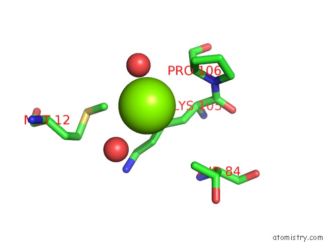
Mono view
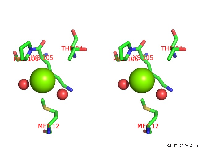
Stereo pair view
Mono view
Stereo pair view
A full contact list of Magnesium with other atoms in the Mg binding
site number 1 of Crystal Structure of Chey Mutant D53A of Helicobacter Pylori within 5.0Å range:
|
Reference:
K.H.Lam,
T.K.Ling,
S.W.Au.
Crystal Structure of Activated CHEY1 From Helicobacter Pylori. J.Bacteriol. V. 192 2324 2010.
ISSN: ISSN 0021-9193
PubMed: 20207758
DOI: 10.1128/JB.00603-09
Page generated: Sun Aug 10 21:52:07 2025
ISSN: ISSN 0021-9193
PubMed: 20207758
DOI: 10.1128/JB.00603-09
Last articles
Mg in 4QV4Mg in 4QV1
Mg in 4QQL
Mg in 4QV3
Mg in 4QUX
Mg in 4QUY
Mg in 4QV0
Mg in 4QTD
Mg in 4QRH
Mg in 4QRE