Magnesium »
PDB 3hd2-3hoz »
3hjf »
Magnesium in PDB 3hjf: Crystal Structure of T. Thermophilus Argonaute E546 Mutant Protein Complexed with Dna Guide Strand and 15-Nt Rna Target Strand
Protein crystallography data
The structure of Crystal Structure of T. Thermophilus Argonaute E546 Mutant Protein Complexed with Dna Guide Strand and 15-Nt Rna Target Strand, PDB code: 3hjf
was solved by
Y.Wang,
H.Li,
G.Sheng,
D.J.Patel,
with X-Ray Crystallography technique. A brief refinement statistics is given in the table below:
| Resolution Low / High (Å) | 43.55 / 3.06 |
| Space group | P 43 21 2 |
| Cell size a, b, c (Å), α, β, γ (°) | 111.868, 111.868, 177.035, 90.00, 90.00, 90.00 |
| R / Rfree (%) | 20.2 / 25.5 |
Magnesium Binding Sites:
The binding sites of Magnesium atom in the Crystal Structure of T. Thermophilus Argonaute E546 Mutant Protein Complexed with Dna Guide Strand and 15-Nt Rna Target Strand
(pdb code 3hjf). This binding sites where shown within
5.0 Angstroms radius around Magnesium atom.
In total only one binding site of Magnesium was determined in the Crystal Structure of T. Thermophilus Argonaute E546 Mutant Protein Complexed with Dna Guide Strand and 15-Nt Rna Target Strand, PDB code: 3hjf:
In total only one binding site of Magnesium was determined in the Crystal Structure of T. Thermophilus Argonaute E546 Mutant Protein Complexed with Dna Guide Strand and 15-Nt Rna Target Strand, PDB code: 3hjf:
Magnesium binding site 1 out of 1 in 3hjf
Go back to
Magnesium binding site 1 out
of 1 in the Crystal Structure of T. Thermophilus Argonaute E546 Mutant Protein Complexed with Dna Guide Strand and 15-Nt Rna Target Strand
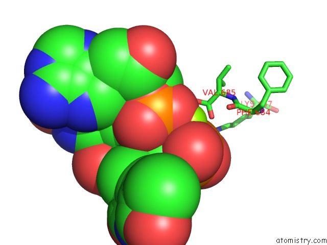
Mono view
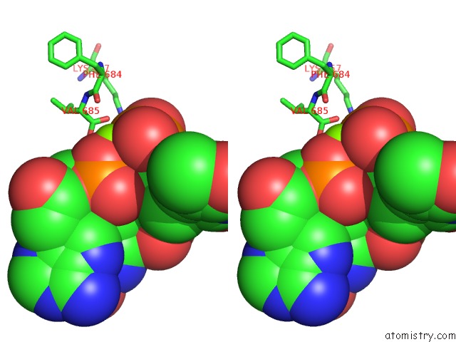
Stereo pair view
Mono view
Stereo pair view
A full contact list of Magnesium with other atoms in the Mg binding
site number 1 of Crystal Structure of T. Thermophilus Argonaute E546 Mutant Protein Complexed with Dna Guide Strand and 15-Nt Rna Target Strand within 5.0Å range:
|
Reference:
Y.Wang,
S.Juranek,
H.Li,
G.Sheng,
G.S.Wardle,
T.Tuschl,
D.J.Patel.
Nucleation, Propagation and Cleavage of Target Rnas in Ago Silencing Complexes. Nature V. 461 754 2009.
ISSN: ISSN 0028-0836
PubMed: 19812667
DOI: 10.1038/NATURE08434
Page generated: Sun Aug 10 21:57:11 2025
ISSN: ISSN 0028-0836
PubMed: 19812667
DOI: 10.1038/NATURE08434
Last articles
Mg in 4JSTMg in 4JS0
Mg in 4JRY
Mg in 4JRC
Mg in 4JRP
Mg in 4JRN
Mg in 4JQP
Mg in 4JR7
Mg in 4JNX
Mg in 4JQL