Magnesium »
PDB 3igi-3is5 »
3ilf »
Magnesium in PDB 3ilf: Crystal Structure of Porphyranase A (Pora) in Complex with Neo- Porphyrotetraose
Protein crystallography data
The structure of Crystal Structure of Porphyranase A (Pora) in Complex with Neo- Porphyrotetraose, PDB code: 3ilf
was solved by
J.H.Hehemann,
G.Correc,
T.Barbeyron,
W.Helbert,
G.Michel,
M.Czjzek,
with X-Ray Crystallography technique. A brief refinement statistics is given in the table below:
| Resolution Low / High (Å) | 49.34 / 1.80 |
| Space group | P 21 21 21 |
| Cell size a, b, c (Å), α, β, γ (°) | 62.090, 68.378, 71.268, 90.00, 90.00, 90.00 |
| R / Rfree (%) | 15.7 / 20.5 |
Other elements in 3ilf:
The structure of Crystal Structure of Porphyranase A (Pora) in Complex with Neo- Porphyrotetraose also contains other interesting chemical elements:
| Chlorine | (Cl) | 2 atoms |
| Calcium | (Ca) | 1 atom |
Magnesium Binding Sites:
The binding sites of Magnesium atom in the Crystal Structure of Porphyranase A (Pora) in Complex with Neo- Porphyrotetraose
(pdb code 3ilf). This binding sites where shown within
5.0 Angstroms radius around Magnesium atom.
In total only one binding site of Magnesium was determined in the Crystal Structure of Porphyranase A (Pora) in Complex with Neo- Porphyrotetraose, PDB code: 3ilf:
In total only one binding site of Magnesium was determined in the Crystal Structure of Porphyranase A (Pora) in Complex with Neo- Porphyrotetraose, PDB code: 3ilf:
Magnesium binding site 1 out of 1 in 3ilf
Go back to
Magnesium binding site 1 out
of 1 in the Crystal Structure of Porphyranase A (Pora) in Complex with Neo- Porphyrotetraose
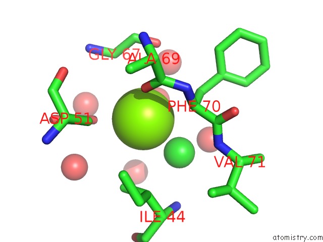
Mono view
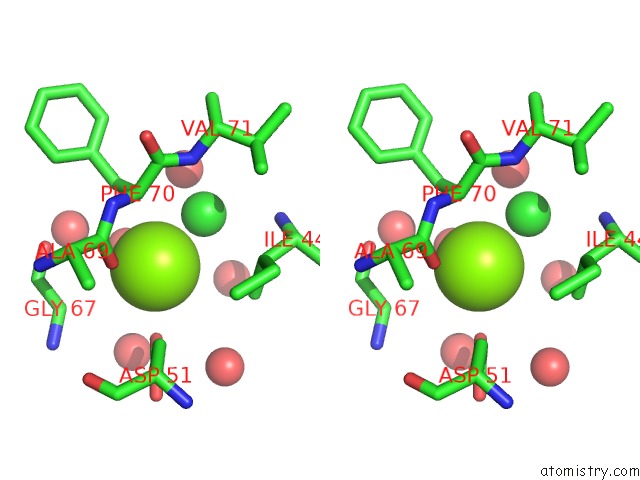
Stereo pair view
Mono view
Stereo pair view
A full contact list of Magnesium with other atoms in the Mg binding
site number 1 of Crystal Structure of Porphyranase A (Pora) in Complex with Neo- Porphyrotetraose within 5.0Å range:
|
Reference:
J.H.Hehemann,
G.Correc,
T.Barbeyron,
W.Helbert,
M.Czjzek,
G.Michel.
Transfer of Carbohydrate-Active Enzymes From Marine Bacteria to Japanese Gut Microbiota. Nature V. 464 908 2010.
ISSN: ISSN 0028-0836
PubMed: 20376150
DOI: 10.1038/NATURE08937
Page generated: Wed Aug 14 16:11:51 2024
ISSN: ISSN 0028-0836
PubMed: 20376150
DOI: 10.1038/NATURE08937
Last articles
Fe in 2YXOFe in 2YRS
Fe in 2YXC
Fe in 2YNM
Fe in 2YVJ
Fe in 2YP1
Fe in 2YU2
Fe in 2YU1
Fe in 2YQB
Fe in 2YOO